
Figures & data
Figure 1. The image pyramid generated by self-similarity.
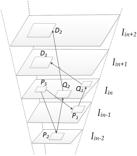
Figure 2. The algorithm flow chart of this paper.
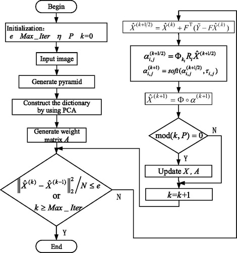
Figure 3. Comparison of the reconstructed images of various methods for noiseless medical image (Equation1(1)
(1) ) (a) Original image (b) LR image (c) Bicubic (d) ASDS (e) Our.
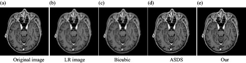
Figure 4. Comparison of the reconstructed images of various methods for noiseless medical image (Equation2(2)
(2) ) (a) Original image (b) LR image (c) Bicubic (d) ASDS (e) Our.
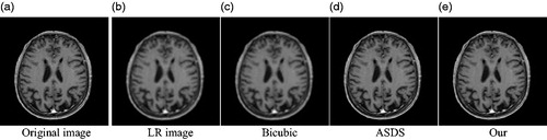
Figure 5. Comparison of the reconstructed images of various methods for noiseless medical image (Equation3(3)
(3) ) (a) Original image (b) LR image (c) Bicubic (d) ASDS (e) Our.
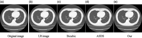
Figure 6. Comparison of the reconstructed images of various methods for noisy medical image (Equation1(1)
(1) ) (a) Original image (b) LR image (c) Bicubic (d) ASDS (e) Our.
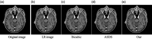
Figure 7. Comparison of the reconstructed images of various methods for noisy medical image (Equation2(2)
(2) ) (a) Original image (b) LR image (c) Bicubic (d) ASDS (e) Our.
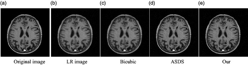
Figure 8. Comparison of the reconstructed images of various methods for noisy medical image (Equation3(3)
(3) ).

Table 1. Comparison of PSNR (dB)/SSIM of the medical images () based on various methods.
Table 2. Comparison of PSNR (dB)/SSIM of the medical images () based on various methods.
Table 3. Comparison of complexity.
Figure 9. Comparison of the reconstructed images of various methods for Butterfly image (a) Original image (b) Bicubic (c) Yang (d) Our.
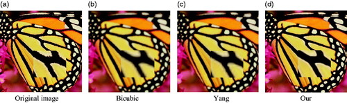
Table 4. Comparison of PSNR (dB)/SSIM of the reconstructed images based on various method.