
Figures & data
Figure 1. (a) The coordinate systems of the surgical drill and the reference. (b) The coordinate systems of the surgical saw and the reference. (c) The calibration for the normal and axis of surgical saw.
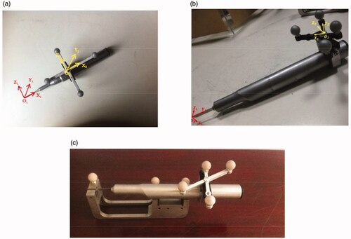
Figure 2. The calibration of the loosed bone graft, and the registration of the whole surgical navigation system.
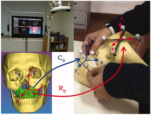
Figure 3. The position and orientation of the surgical saw were tracked in real time and displayed on the computer screen so that the maxilla was resected according to the preoperative planned trajectory.
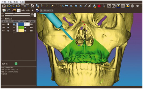
Figure 4. The reposition of the loosed maxilla under the interactive guidance of 2D and 3D image rendering environment. The red and green contours in the 2D views respectively represent the positions of the intraoperative tracked and the preoperative planned loosed maxilla. The red and green models in the 3D view, respectively, represent the intraoperative tracked and the preoperative planned loosed maxilla.
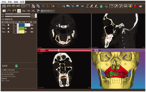
Figure 5. The error distribution of the loosed maxilla reposition. (a) Non-navigated (free-hand operation) reposition. (b) Image-guided reposition.
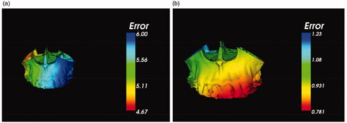
Table 1. Group 1: The distance error of homologous points on the preoperative planned and the non-navigated (free-hand operation) reposition models. Group 2: The distance error of homologous points on the preoperative planned and the image-guided reposition models.