Figures & data
Figure 1. The appearance of the patient’s right ankle. The ankle was swollen (a, b) and distorted (c). The skin on the back of right foot (b) was scared seriously because of one fire accident before.
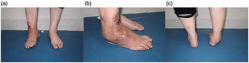
Figure 2. Radiological views in right ankle at anteroposterior (left), lateral (right) position (a) and 3D reconstruction of CT scanning in CN ankle (b).
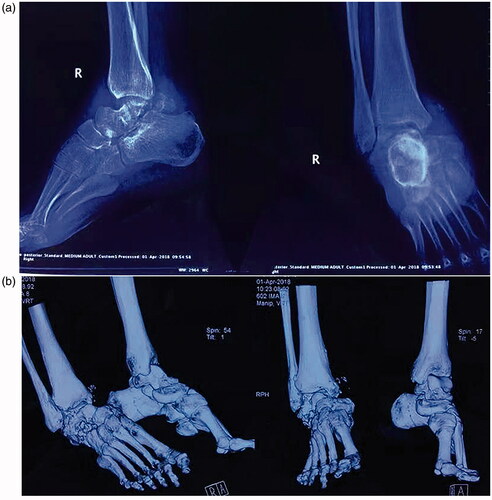
Figure 3. The design of titanium implant for 3DP based on the results of 3D reconstruction of CT scanning (a–c). The preoperative planning by computer assistance for the placement of Retrograde interlocking intramedullary nail (d).
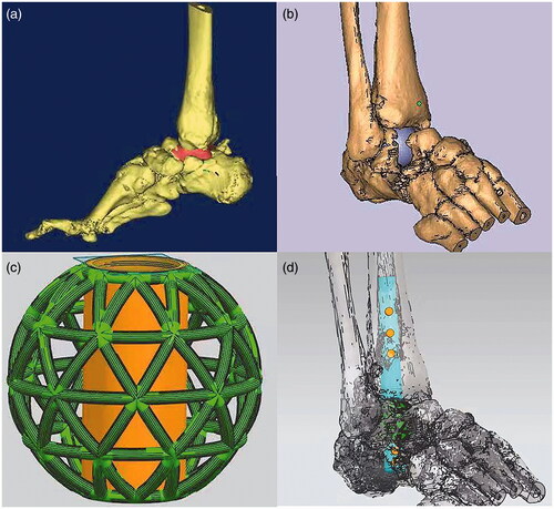
Figure 4. Surgical process in right ankle of patient with CN (a–c, e, f). Initially two sizes, diameter 3 and 3.4 cm respectively, of metal implant (d) produced by manufacture. In practice, the bigger one was implanted in the anatomic place of talus.
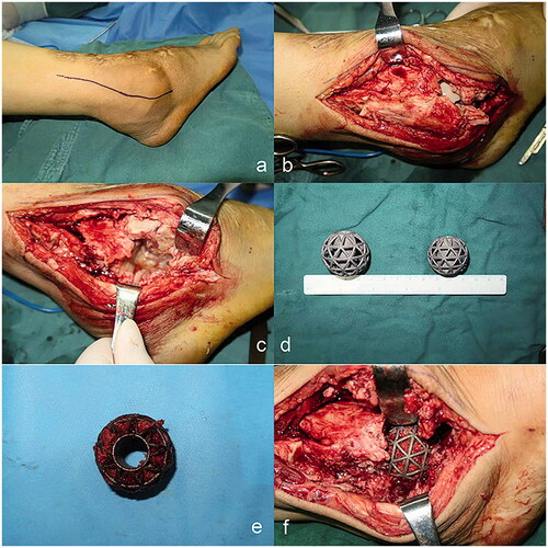
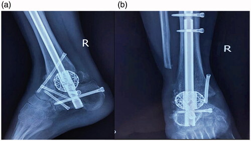
Figure 5. No signs of dislocation, nonunion, pathologic fractures or pathological bone resorption were shown in right ankle by a/p (a) and lateral (b) radiographic evaluation at 3 months follow-up.