Figures & data
Table 1. Summary of technical approaches for neoadjuvant studies.
Table 2. Summary of technical approaches for adjuvant studies.
Table 3. Performance metrics from neoadjuvant studies in this review.
Table 4. Performance metrics from adjuvant studies in this review.

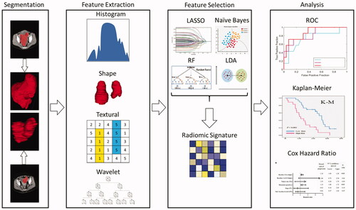
![Figure 1. This graphic gives a patient a top-level view of their progression from an initial screening, chemotherapy, surgery, and surveillance after resection. Radiomics serve as a diagnostic and prognostic tool, which can be applied at many different timepoints in oncologic care. Use of this technology in the diagnosis stage, treatment planning stage, and surveillance stage can complement traditional oncologic care and help personalize treatment options for patients with solid tumors [Citation23].](/cms/asset/8bb2032b-511e-4e05-bbe5-938d387af85f/icsu_a_1994014_f0001_c.jpg)
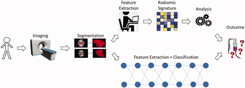
![Figure 2. This flowchart shows the generalized overview of the neoadjuvant and adjuvant studies from patient image acquisition (CT, MRI, PET), tumor segmentation, radiomic signature, machine learning analysis, and the predicted outcome. These studies capture prognosis, recurrence, therapeutic response, survival, and tumor volume. These various outcome categories describe different aspects of the effectiveness of either neoadjuvant or adjuvant chemotherapy [Citation24,Citation25].](/cms/asset/2f8676dc-ec5c-4d69-a9ed-45e31fad8327/icsu_a_1994014_f0002_c.jpg)