
Figures & data
Figure 1. Components of the navigation system: a tablet device (iPad 2® Apple Inc., CA, USA), CT-compatible markers, and plastic marker caps.

Figure 2. Screens during surgical navigation: the yellow object indicates a vascular structure. (a) Preoperative view of the tablet using a small triangle. Red and yellow crosses indicate perforator positions at the level of emergence from the fascia; (b) preoperative view of the tablet using a large triangle; (c) a 3D perforator image using a 3D slicer; (d) intraoperative view of the navigation system.
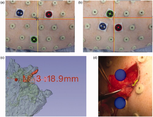
Figure 3. 3D vascular structures based on CT images. Based on the data, we selected perforators to be used in our experiment. (a) Lateral view; (b) dorsal view.
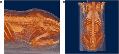
Table 1. Errors of tablet device navigation system registered by small and large triangles.
Table 2. Coordination measured by optical tracking system (mm).