
Figures & data
Figure 1. Lateral scalloping with macroglossia.
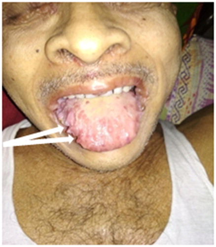
Figure 2. Papules and plaque over periocular, perinasal and perioral area.
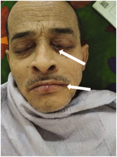
Figure 3. Anterior shoulder pad sign.
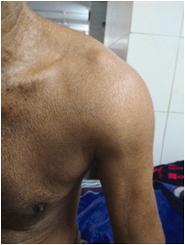
Figure 4. Soft tissue swelling over the wrist due to amyloid deposit.
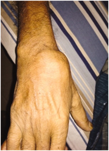
Figure 5. (a) and (b) Plaque biopsy shows amyloid deposit confirmed by Congo red staining with apple green birefringence.
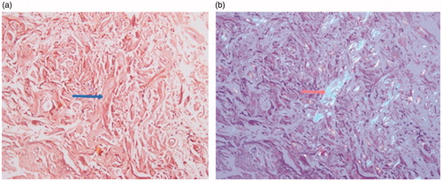
Figure 6. High-power appearance of the spread bone marrow aspirate showing normal bone particles of bone marrow at the end of cellular trails.
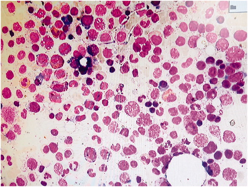
Figure 7. Increased accumulation of Lambda light chain in the Lambda zone on serum IFE.
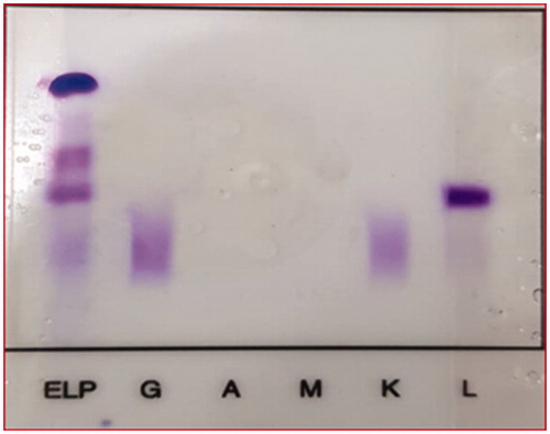
Table 1. The serum free light chain (FLC) assay.