Figures & data
Figure 1. Brain MRI axial imaging slides showing diffuse cortical T2-weighted hyperintensities, symmetrical T2-weighted hyperintensities in the midbrain and hippocampi (panel A), basal nuclei, and thalami (panel B). MRI coronal imaging slides show basal ganglia, thalami, and hippocampi involvement (panel C) with extension into midbrain, pons, and medulla (panel D).
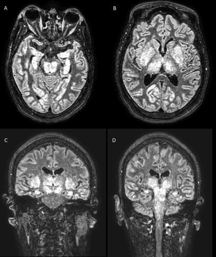
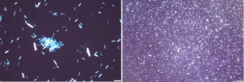
Figure 2. Urine microscopic evaluation showed numerous needle-shape polarizable crystals, morphologically compatible with calcium oxalate monohydrate crystals (Panel A 40X, Panel B 4X).