
Figures & data
Figure 1. (a) x-ray of right elbow showing significant joint degeneration. (b) MRI of the spine showing superior end plate compression fractures in T12 and L1 with minimal loss of height. (c) Bone scan revealing widespread increased uptake
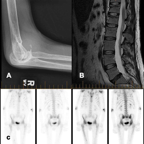
Table 1. Etiologies of pain in patients with Hemophilia
Supplemental material