Figures & data
Figure 1. Hematoxylin and eosin staining of paraffin sections of an Erpobdella johanssoni ovary. A, Histological sections of the ovarian cord from the control group showing normal architecture: nurse cells (NC) and vitellogenic oocytes (VO). B, Ovary from treated leech revealed degeneration shrinkage of nurse cells (NC) and vitellogenic oocytes (VO), and extensive vacuolization of their ooplasm (arrowheads). Scale bars = 20 μm.
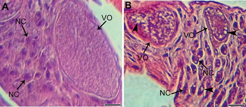
Figure 2. A, Ultrastructure of asomatic cell and vitellogenic oocyte in ovarian cord from control. During oogenesis, the ooplasm gathers cytoplasmic organelles, mainly mitochondria (MI), reserve material and microvilli (MV). B, An overview of a somatic cell undergoing the initial stages of cell death caused by cytoplasmic vacuolization and formation of autophagic vacuole (large arrow), chromatin condensation with dense clumps abutting nuclear membrane, degenerating mitochondria (MI), and the loss of their lamellar cristae. C, Vitellogenic oocytes showing degenerated cytoplasm, damage of microvilli (MV) in some zones, and damage to the vitelline envelope (VE) in others. N: nucleus; SC: somatic cell; OO: ooplasm. Scale bars: A = 5 µm; B = 1 µm; C = 2 µm.
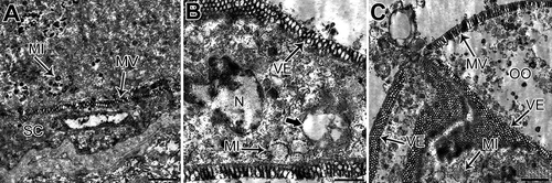
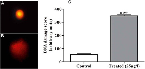
Figure 3. A, B, Score of DNA damage in ovarian cells showing a higher score of DNA alteration in 25 µg L−1 treatment than in the control group. C, Values expressed as the mean ± standard deviation (SD) from at least three separate experiments. (*) p < 0.05 indicates significant difference from control (***) p < 0.0001.