Figures & data
Figure 1. Map of the study area and location of sampling stations (St.).
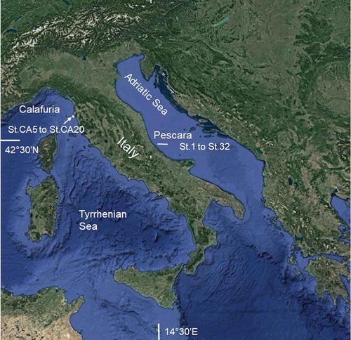
Table I. Details, water parameters at the bottom, TOC (total organic carbon) concentrations in sediment and sediment granulometric composition of the stations where specimens of Chaetozone corona Berkeley & Berkeley, Citation1941 and C. carpenteri McIntosh, Citation1911 were recorded in the Central Adriatic Sea and Tyrrhenian Sea. n.e. = not examined.
Figure 2. Chaetozone corona. (a) Dorsal view, anterior end, with dorsal tentacle (dt) and branchiae (br). (b) Dorsal view of the notopodia showing spines and capillary chaetae, middle part of the body. (c) Posterior part of the body and pygidium, dorso-lateral view. Scale bars: a = 100 µm; b = 30 µm; c = 20 µm.
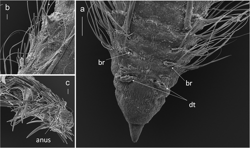
Figure 3. Occurrence of Chaetozone corona Berkeley & Berkeley, Citation1941 and C. carpenteri McIntosh, Citation1911 in the Central Adriatic Sea: densities (average individuals m−2) ± standard error.
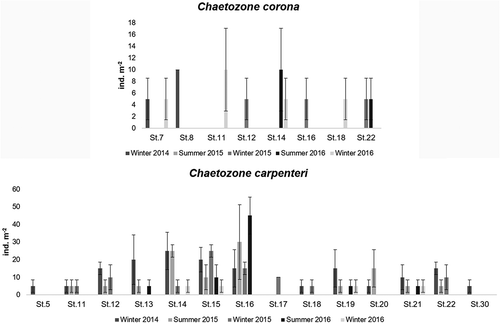
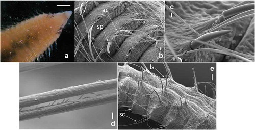
Figure 4. Chaetozone carpenteri. (a) Dorsal view of prostomium showing eyes and black speckles, anterior end. (b) Lateral view of awl-shaped capillary (ac) chaetae and large spines (sp), anterior end. (c) Detail of large spine. (d) Detail of damaged chaetae of first chaetigers. (e) Lateral view of long spines (ls) and short capillaries (sc), posterior end. Scale bars: a = 0.5 mm; b = 20 µm; c = 10 µm; d = 2 µm; ee = 100 µm.