1,091
Views
4
CrossRef citations to date
0
Altmetric
Articles

Effects of X-ray irradiation on haemocytes of Procambarus clarkii (Arthropoda: Decapoda) males

, ORCID Icon, , , , , & show all
Pages 26-35 | Received 29 Sep 2017, Accepted 20 Dec 2017, Published online: 22 Jan 2018

Figures & data

Figure 1. Transmission electron microscopy of Procambarus clarkii haemocytes, CTRL group. (a,b) Hyaline haemocytes (HH) showing a high nucleus/cell ratio. (c) Granular (GH) and semigranular haemocytes (SH). Transversal (d,e) and longitudinal (f) section of granular haemocytes (GH). Arrows: granules; ly: lysosome; m: mithocondria; n: nucleus; rer: rough endoplasmic reticulum. Scale bars: a, b, d–f = 2 µm; c = 5 µm.
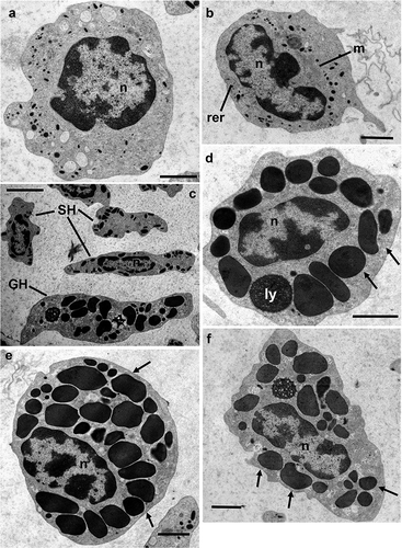
Figure 1. Transmission electron microscopy of Procambarus clarkii haemocytes, CTRL group. (a,b) Hyaline haemocytes (HH) showing a high nucleus/cell ratio. (c) Granular (GH) and semigranular haemocytes (SH). Transversal (d,e) and longitudinal (f) section of granular haemocytes (GH). Arrows: granules; ly: lysosome; m: mithocondria; n: nucleus; rer: rough endoplasmic reticulum. Scale bars: a, b, d–f = 2 µm; c = 5 µm.

Figure 2. Transmission electron microscopy of Procambarus clarkii haemocytes, latex beads group. (a,b) semigranular haemocyte (SH). (c,d) medium granules haemocyte (MH). (e,f) Semigranular haemocyte (SH) involved in phagocytic activity after in vivo artificial non-self-challenge. A large number of latex beads (asterisks) are present in the cytoplasm included into the phagosomes. (g) Detail of semigranular haemocytes (SHs) showing the latex bead phagocytosis at the membrane level. Many granules appear to be fusing with phagosomes (arrowheads). n: nucleus. Scale bars: b = 1 µm; a, c–f = 2 µm; g = 500 nm.
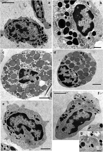
Figure 2. Transmission electron microscopy of Procambarus clarkii haemocytes, latex beads group. (a,b) semigranular haemocyte (SH). (c,d) medium granules haemocyte (MH). (e,f) Semigranular haemocyte (SH) involved in phagocytic activity after in vivo artificial non-self-challenge. A large number of latex beads (asterisks) are present in the cytoplasm included into the phagosomes. (g) Detail of semigranular haemocytes (SHs) showing the latex bead phagocytosis at the membrane level. Many granules appear to be fusing with phagosomes (arrowheads). n: nucleus. Scale bars: b = 1 µm; a, c–f = 2 µm; g = 500 nm.

Figure 3. Diameter (a) and circularity (b) of granules characterising the four types of haemocytes. CTRL: control group, HH: Hyaline haemocytes, SH: semigranular haemocytes, MH: medium granule haemocytes, GH: granular haemocytes. IRR: irradiated group.
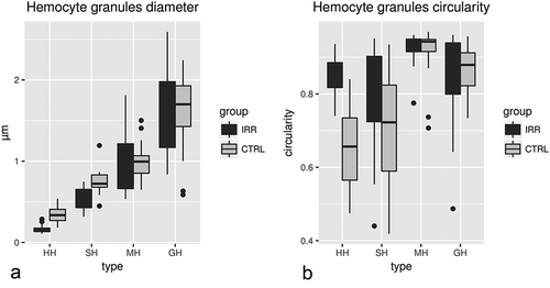
Figure 3. Diameter (a) and circularity (b) of granules characterising the four types of haemocytes. CTRL: control group, HH: Hyaline haemocytes, SH: semigranular haemocytes, MH: medium granule haemocytes, GH: granular haemocytes. IRR: irradiated group.

Figure 4. Transmission electron microscopy of Procambarus clarkii haemocytes 20 days after irradiation at a dose of 40 Gy. (a) Hyaline haemocytes: HH, Hyaline haemocyte. (b) Semigranular haemocytes: SH, Semigranular haemocyte. (c) Granular haemocytes: GH, Granular haemocyte. (d,e) Medium granule haemocytes: MH. (f) Detail of structured granules in MH, Medium granule haemocyte. (g) GH-like granules, sg: structured granules. Scale bars: f = 1 µm; a, b, d, e = 2 µm; c = 5 µm.
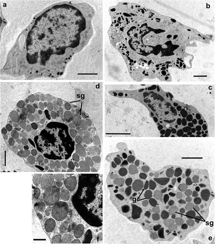
Figure 4. Transmission electron microscopy of Procambarus clarkii haemocytes 20 days after irradiation at a dose of 40 Gy. (a) Hyaline haemocytes: HH, Hyaline haemocyte. (b) Semigranular haemocytes: SH, Semigranular haemocyte. (c) Granular haemocytes: GH, Granular haemocyte. (d,e) Medium granule haemocytes: MH. (f) Detail of structured granules in MH, Medium granule haemocyte. (g) GH-like granules, sg: structured granules. Scale bars: f = 1 µm; a, b, d, e = 2 µm; c = 5 µm.

Figure 5. Total circulating haemocytes from control (CTRL), PBS group (PBS), latex beads (LB) and irradiated (IRR) groups.
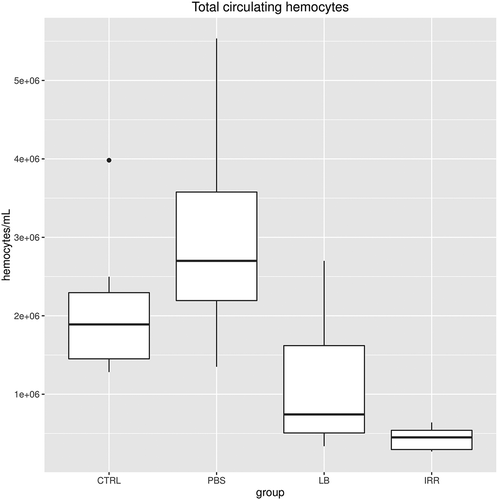
Figure 5. Total circulating haemocytes from control (CTRL), PBS group (PBS), latex beads (LB) and irradiated (IRR) groups.

Figure 6. Differential haemocyte percentages in Procambarus clarkii male from control (CTRL), PBS group (PBS), latex beads (LB) and irradiated (IRR) groups.
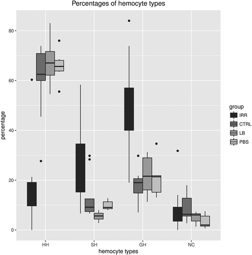
Figure 6. Differential haemocyte percentages in Procambarus clarkii male from control (CTRL), PBS group (PBS), latex beads (LB) and irradiated (IRR) groups.

Figure 7. Basal (PO) and total plasmatic phenoloxidase (pPO) activities in Procambarus clarkii males from control (CTRL), latex beads (LB) and irradiated (IRR) groups measured as the slope of the reaction curve at Vmax. The enzymatic activities were recorded as absorbance units for µL of haemolymph per min (for statistics see the text).
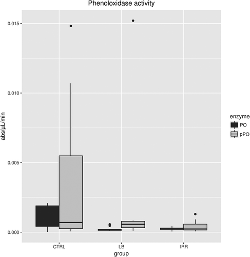
Figure 7. Basal (PO) and total plasmatic phenoloxidase (pPO) activities in Procambarus clarkii males from control (CTRL), latex beads (LB) and irradiated (IRR) groups measured as the slope of the reaction curve at Vmax. The enzymatic activities were recorded as absorbance units for µL of haemolymph per min (for statistics see the text).