Figures & data
Figure 1. Vincenzo Briganti. Vincenzo [or Vincenzio] Briganti (1766–1836). Unedited photo by Iconoteca dei Botanici, Library of Orto Botanico, Università degli Studi di Padova. Arrangement: IB.NN.26
![Figure 1. Vincenzo Briganti. Vincenzo [or Vincenzio] Briganti (1766–1836). Unedited photo by Iconoteca dei Botanici, Library of Orto Botanico, Università degli Studi di Padova. Arrangement: IB.NN.26](/cms/asset/74f523fd-c1d2-4359-a64d-ce5d9ce97cbb/tizo_a_1892217_f0001_oc.jpg)
Table I. Specimens of H. straminea of the J. R. Bourguignat Collection of the MHNG
Table II. Comparison of anatomical and morphological data (in mm) in H. mileti (4 specimens: Posta, Rieti, Latium, 715 m a.s.l.; Gallo Matese, Caserta, Campania, 830 m a.s.l.; Mount Miletto, San Massimo, Campobasso, 1600 m a.s.l. and Oratino, Campobasso, 360 m a.s.l., Molise), H. straminea (4 specimens: Laviano, 600 m a.s.l.; Pietracamela, Teramo, 1050 m a.s.l. and Padula, Cortino, Teramo, 950 m a.s.l., Abruzzo; Oratino, Campobasso, Molise, 360 m a.s.l.) and H. lucorum (12 specimens, 3 per station: Govone, Cuneo, 237 m a.s.l.; Cà Conti, Granze, Padova, 6 m a.s.l.; Mandriole, Ravenna, 4 m a.s.l.; Jesolo, Venezia, 1 m a.s.l.)
Figure 2. Original plate of Briganti. The original by Briganti (Citation1825) showing the shell of two examples of “Elice straminea” () and of the variety γ “shell with almost nil stramineous coloured bands () (Photo by G. Di Dato)
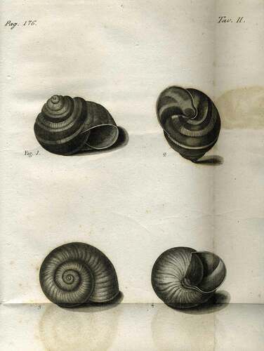
Figure 3. Original plate of Gualtieri. (a) Cochlea terrestris vulgaris, rufescens, fasciata (figure B, plate 1) of Gualtieri (Citation1742) cited by Briganti (Citation1825). (b) Cochlea terrestris vulgaris, cinerea, aliquando pulla, fasciis quatuor fulvis distincta (figure C, plate 1) of Gualtieri (Citation1742)
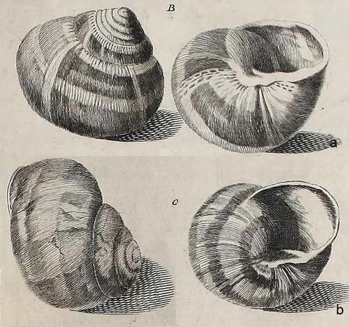
Figure 4. Original plate of Bourguignat. The Table 20, by Bourguignat (Citation1860b)
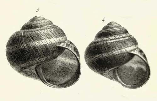
Figure 5. Original plate of Kobelt. Figure 1027, var. anaphora coming from Bari (Kobelt Citation1876)
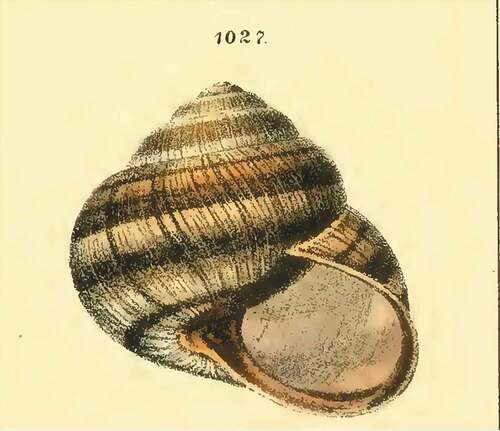
Figure 6. Lectotype of Helix straminea Briganti, Citation1825. (a) Original sample of V. Briganti catalogued n. MHNG-MOLL-118135, collection of J.R. Bourguignat, Muséum d’Histoire Naturelle of Genève, herein designated as lectotype of Helix straminea V. Briganti Citation1825. (b) Original anonymous label not autograph. (c,d) Details with other labels. (e) Associated autograph label of Bourguignat (Photos by E. Tardy)
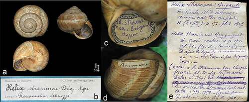
Figure 7. Paralectotype of Helix straminea Briganti, Citation1825. Original sample of V. Briganti catalogued n. MHNG-MOLL-118136, collection of J.R. Bourguignat, Muséum d’Histoire Naturelle of Genève, here designated as paralectotype of Helix straminea V. Briganti Citation1825. (Photos by E. Tardy)
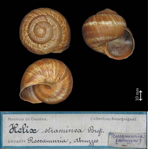
Figure 8. Specimen of Bourguignat collection. (a) Specimen no. MHNG-MOLL-118129 of Bourguignat collection. (b) Outside original Label. (c) Inside original label with a different calligraphy from that of Bourguignat (Photos by E. Tardy). (d) Original label of a specimen of H. straminea from Paulucci collection n. MZUF-1698, collected by I. Blanc in 1877 (Photos by S. Cianfanelli). The calligraphy of the two labels clearly corresponds
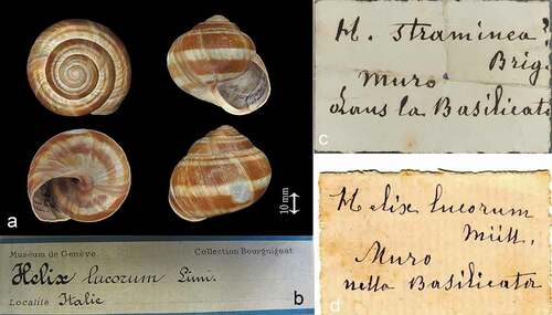
Figure 9. Giemsa stained karyotype of the sample from Ruvo del Monte (Potenza, Italy) (Photo by G. Odierna)
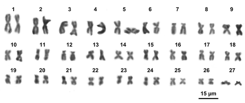
Figure 10. Metaphase plate of H. straminea from Ruvo del Monte sequentially stained with C-banding+Giemsa (a), CMA (b) and Dapi (c) (Photo by G. Odierna)
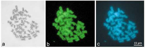
Figure 11. MRBAYES consensus 16S+COI tree of Helix straminea. Numbers at nodes are posterior probabilities
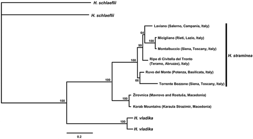
Figure 12. Reproductive system of Helix straminea from Laviano (Salerno). (a) Complete genitalia. (b) Distal genitalia and internal structure of penis and vagina. (c) Proximal penis section. (d) Dart and dart section. (e) Mantle edge and body lobes. (f) Jaw. a = anus, ag = albumen gland, bc = bursa copulatrix, bcd = duct of the bursa copulatrix, bw = body wall, dg = digital gland, do = dart opening, dp = distal penis, ds = dart sac, dv = distal vagina, e = epiphallus, f = flagellum, fhd = first hermaphrodite duct, fo = free oviduct, ga = genital atrium, ldl = left dorsal lobe, lll = left lateral lobe, o = ovotestis, po = pneumostomal opening, pp = proximal penis, ppo = proximal penis opening, pos = prostatic ovispermiduct, pr = penis retractor muscle, ps = penial sheat, pv = proximal vagina, rl = right lobe, sl = subpneumostomal lobe, t = talon, uos = uterine ovispermiduct, v = verge, vd = vas deferens. (Draft by I. Niero)
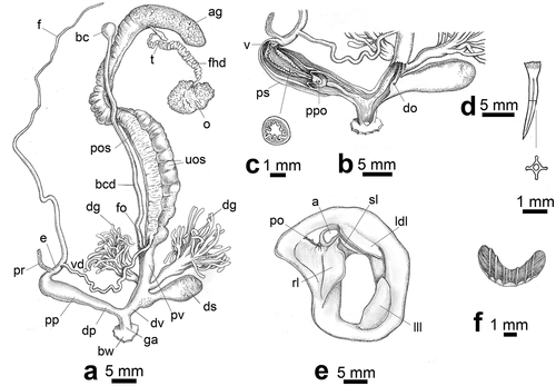
Figure 13. Protoconch development. (a) Helix lucorum. (b) Helix mileti. (c) Helix straminea. (Scale bar on the right 1 mm)
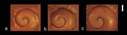
Figure 14. SEM images of radula of H. straminea. (a) Anterior-posterior (from top to bottom) middle section of the radula. (b) Detail. (C) Central and first two lateral teeth. (d) Central tooth. (e) First right lateral tooth. (f) First left lateral tooth. (g) Outermost lateral teeth. (h) Detail of the outermost lateral tooth. (i) Marginal teeth. (j) Detail of a marginal tooth. (k) Lateral teeth seen from the outer edge. (l) Lateral teeth of the anterior part of the radula clearly worn. (m) Detail. (n) An anterior lateral teeth clearly worn. (o) A thin film of dehydrated mucous that covers a tooth. (p) Detail of the film. (Photos by S. Sorbo)
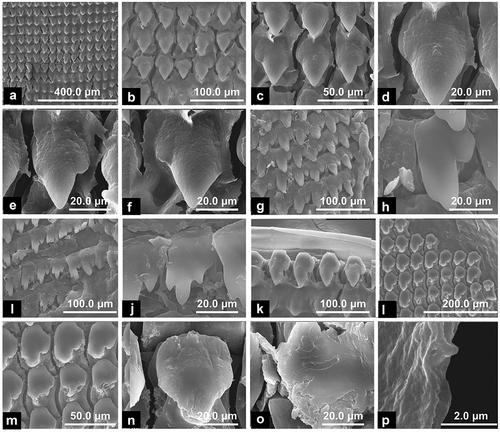
Figure 15. Lectotype of Helix straminiformis Bourguignat, Citation1876. Samples no. MHNG-MOLL-118064 from “Mont Amaro, Abruzzes” of Bourguignat collection, here designated as lectotype of Helix straminiformis Bourguignat, Citation1876. (Photo by E. Neubert & E. Bochud)
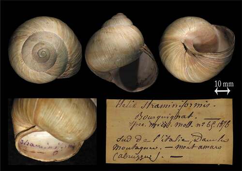
Figure 16. Lectotype of Helix yleobia Bourguignat, Citation1883. (a) Specimen no. MHNG-MOLL-118139 from Tricarico of Bourguignat collection, here designated as lectotype of Helix yleobia Bourguignat, Citation1883. (b) Original label with a calligraphy ascribable to Ippolito Blanc of Nice. (c) Inside label. (d,e) Other labels associated with the specimen. (Photo by E. Neubert & E. Bochud)
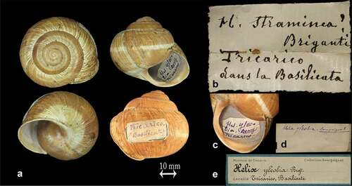
Figure 17. Lectotype of Helix straminea elongata Bourguignat, 1860. (a) Specimen catalogued under No. MHNG-MOLL-118145-1, Bourguignat collection, not taken into account for the typical series. (b) Original label of lot. (c) Associated autograph label of Bourguignat. (d) Specimens catalogued under No. MHNG-MOLL-118145-2 (4), here designated as lectotype of Helix straminea ssp. elongata Bourguignat, 1860. (e) Detail. (Photos by E. Tardy)
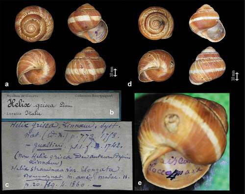
Figure 18. Comparison between Bourguignat figures and specimens present in MHNG. (a) Original figure of H. straminea var. elongata, , pl. 20 (Bourguignat Citation1860b). (b) Specimen no. MHNG-MOLL-118145-2 (4). (c) Specimen no. MHNG-MOLL-118135. (d) Original figure of Bourguignat (Citation1860b) , pl. 20 of H. straminea. (e) Specimen no. MHNG-MOLL-118136
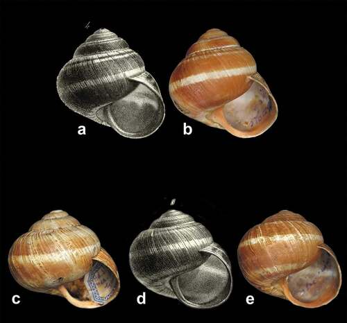
Figure 19. Samples of Shuttleworth collection in the NMBE. Samples collected by C. Sury from the Robert James Shuttleworth collection, preserved in the NMBE, respectively from Capri and Naples. (Photos by O. Korábek). (a) NMBE512977/4. (b) NMBE512978/6
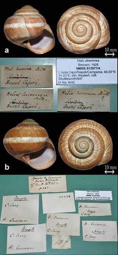
Figure 20. Some of 18 specimens of Helix asemnis Bourguignat, 1860 preserved in the collections of MANN (Photos by N. Maio)

Table III. Specimens of H. straminea of the museum collections with interesting faunistic data
Figure 21. Living specimen of H. straminea from South of Italy. (a–c) A living specimen found in Laviano (Province of Salerno) in 2015. (d–f) Shell of a specimen from Laviano. (g–i) A living specimen found in Ruvo del Monte (Province of Potenza) in 2018. (Photos by N. Maio)
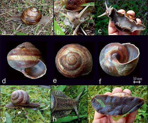
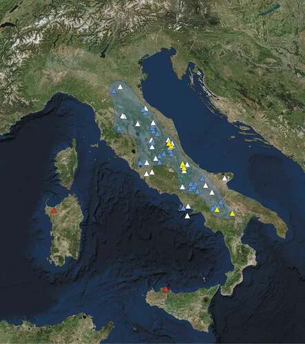
Figure 22. Distribution of Helix straminea in Italy. The updated extent of occurrence (EOO) (bluish area). Historical data before 1980 (white triangles); recent published and new unpublished records (azure triangles). Type localities of the species and synonyms are indicated by yellow triangles. Localities where the species is introduced or accidental (red triangles). ?Doubtful data. (Picture by ESRI satellite, free edition, 2018, modified by S. Viglietti)