Figures & data
Figure 1. (a) Study area, boxes encircling the sampling site, previous occurrences of Styela clava in the Mediterranean Sea are numbered in temporal order; (b) alive specimens of S. clava collected in Lagoon of Venice.
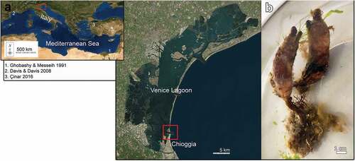
Figure 2. Styela clava. (a) Individual of S. clava with the characteristic leathery and mammillated brown tunic and a long dark brown stalk (asterisk) and the siphons lying close to each other (arrow); (b–c) specimens without tunic showing the gonads; (b) the left side, testicular follicles constituted by small white lobes (ts) separated from the long pinkish female gland (oo), siphons marked with brown stripes (arrow); (c) right side; (d) dissected specimens without branchial sac showing gonads on both sides; (e) magnification of the gonads showing the endocarps (e), testis (ts) and ovaries (oo). Scale bars: 1 cm.
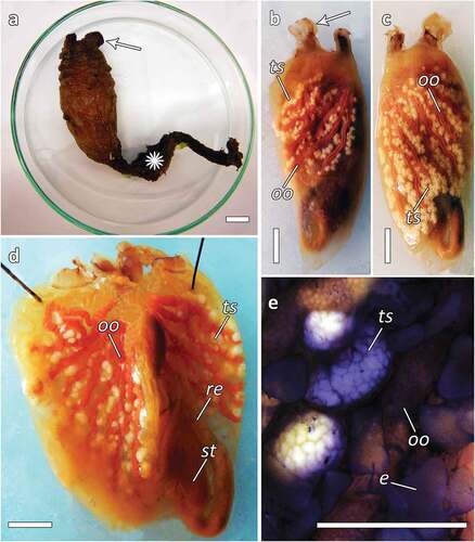
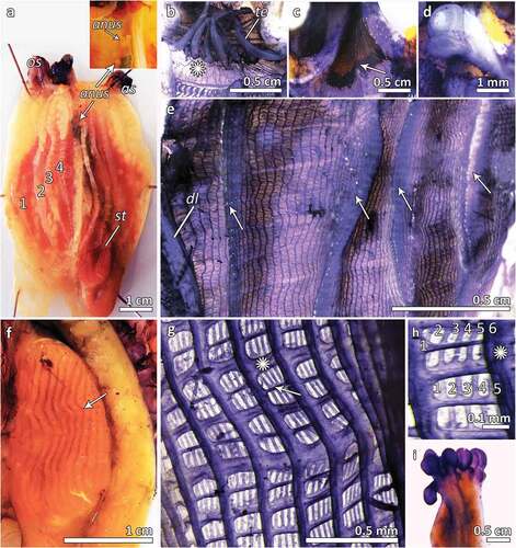
Figure 3. (a) Dissected specimen of S. clava showing the branchial sac with four folds per side, the oral siphon (os) and the atrial one (as), the stomach (st) and the anus opening (anus), with magnification of the anus (anus); (b) oral siphon with long thick oral tentacles (te) and smooth prebranchial area (asterisk); (c) atrial siphon without tentacles (arrow); (d) dorsal tubercle spiral-shaped; (e) branchial sac showing four folds from the dorsal lamina (dl), and 12 to 44 longitudinal vessels on the 4 branchial folds and from 4 to 20 in the interspace; (f) stomach with about 20 folds per side (arrow); (g) branchial sac with transverse vessels of two different size (asterisk points out a thicker one) (h); branchial mesh with about 5–6 stigmata, the asterisk points out the longitudinal vessel; (i) lobed anus.