
Figures & data
Figure 1. Microstructure of femoral bone in brown bears cubs. 1: non-vascular bone tissue, 2: dense Haversian bone tissue, 3: plexiform bone tissue with isolated secondary osteons, orange arrows: lines of arrested growth (LAGs).
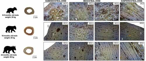
Figure 2. Microstructure of femoral bone in adult brown bears. 1: non-vascular bone tissue, 2: dense Haversian bone tissue, 3: plexiform bone tissue with isolated secondary osteons, orange arrows: lines of arrested growth (LAGs).
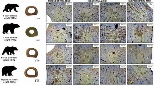
Table I. Schematic histomorphological data on the bear compact bone tissue
Table II. Morphometrical results of brown bears compact bone tissue
Figure 3. Scatter plots showing the correlation between mean osteon diameter and mean Haversian canal diameter both in cubs and adult bears. Values are expressed in μm.
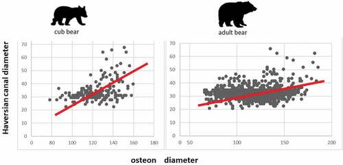
Table III. Data of the eccentricity (e) of osteons and Haversian canals in brown bears
Table IV. Statistical data on the variance of osteon diameter, Pearson’s correlation, and regression-line functions of the correlation between the diameter of osteons and Haversian canals