
Figures & data
Table I. Collecting site locations, characteristics and collecting dates for new species. Abbreviations: j – juvenile, l – larva, u – sex unidentified. The geographic reference system: WGS84.
Table II. GenBank accession numbers for the Echiniscoides spp. sequenced in this work.
Figure 1. Bayesian phylogeny of Echiniscoididae based on COI. Values at the nodes signify posterior probabilities. Echiniscus testudo was used as an outgroup. The scale bar represents 0.5 substitutions per nucleotide position.

Table III. Measurements [in µm] of selected morphological structures of individuals of Echiniscoides basalticus sp. nov. (type series) mounted in Hoyer’s medium. Abbreviations: N – number of females/structures measured, Range refers to the smallest and the largest structure among all measured specimens; SD – standard deviation.
Figure 2. Habitus of Echiniscoides basalticus sp. nov. (PCM): A. holotypic female, B. allotypic male. List of abbreviations (identical for all figures): cA – cirrus A, ce – cirrus externus, cE – cirrus E, ci – cirrus internus, p1–4 – sense organs on legs I–IV, pc – primary clava, sc – secondary clava (cephalic papilla). Scale bars in μm.
Figure 3. Drawing of Echiniscoides basalticus sp. nov. Scale bar in μm.
Figure 4. Buccal apparatus of Echiniscoides basalticus sp. nov. (PCM). Scale bar in μm.
Table IV. Measurements [in µm] of selected morphological structures of adult females of Echiniscoides bufocephalus sp. nov. (type series) mounted in Hoyer’s medium. Abbreviations: N – number of specimens/structures measured, Range refers to the smallest and the largest structure among all measured specimens; SD – standard deviation.
Figure 5. Habitus of the holotypic female of Echiniscoides bufocephalus sp. nov. (PCM). Scale bar in μm.
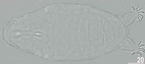
Figure 6. Drawing of Echiniscoides bufocephalus sp. nov. Scale bar in μm.
Figure 7. Detailed morphology of Echiniscoides bufocephalus sp. nov.: A. specimen in toto (SEM), B. head in close-up (dorsal view, SEM), C. head in close-up (ventral view, SEM), D. buccal apparatus (PCM). Scale bars in μm.
Figure 8. Cephalic and trunk appendages of Echiniscoides bufocephalus sp. nov.: A. peribuccal cirri and secondary clava (PCM), B. cirrus internus in close-up (SEM), C. cirrus A and primary clava (SEM), D. sensory organ on leg II (SEM), E. sensory organ on leg III (PCM), F. cirrus E (SEM). Scale bars in μm.
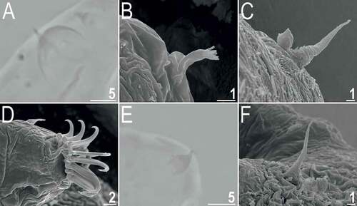
Figure 9. Detailed morphology of Echiniscoides bufocephalus sp. nov.: A. claws I (PCM), B. claws IV (SEM), C. anus and male gonopore (SEM), D. male gonopore in close-up (SEM). Scale bars in μm.
Table V. Measurements [in µm] of selected morphological structures of adult females of Echiniscoides lichenophilus sp. nov. (type series) mounted in Hoyer’s medium. Abbreviations: N – number of specimens/structures measured, Range refers to the smallest and the largest structure among all measured specimens; SD – standard deviation.
Table VI. Measurements [in µm] of selected morphological structures of adult males of Echiniscoides lichenophilus sp. nov. (type series) mounted in Hoyer’s medium. Abbreviations: N – number of specimens/structures measured, Range refers to the smallest and the largest structure among all measured specimens; SD – standard deviation.
Figure 10. Habitus of the holotypic female of Echiniscoides lichenophilus sp. nov. (PCM). Scale bar in μm.
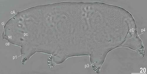
Figure 11. Drawing of Echiniscoides lichenophilus sp. nov. Scale bar in μm.
Figure 12. Detailed morphology of Echiniscoides lichenophilus sp. nov. (DIC): A. buccal apparatus, B. caudal body portion with cirri E, C. claws II and sense organ on leg II. Scale bars in μm.
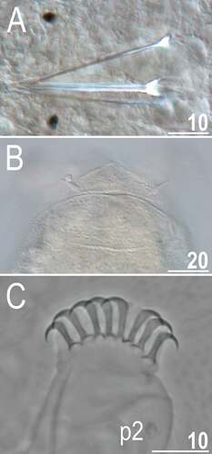
Figure 13. Detailed morphology of Echiniscoides lichenophilus sp. nov. (SEM): A. specimen in toto, B. cephalic body portion with peribuccal cirri in frontal view, C. cephalic body portion with peribuccal cirri in lateral view, D. cirrus A and primary clava, E. cirrus E and sense organ on leg IV, F. claws II, G. claws III. Scale bars in μm.
Table VII. Measurements [in µm] of selected morphological structures of adults of Echiniscoides musa sp. nov. (type series) mounted in Hoyer’s medium. Abbreviations: SD – standard deviation.
Figure 14. Detailed morphology of Echiniscoides musa sp. nov. (PCM): A. holotypic female, B. anterolateral body portion of paratypic female, C. buccal apparatus, D. claws II. Scale bars in μm.
Figure 15. Drawing of Echiniscoides musa sp. nov. Scale bar in μm.
Table VIII. Measurements [in µm] of selected morphological structures of adult females of Echiniscoides trichosus sp. nov. (type series) mounted in Hoyer’s medium. Abbreviations: N – number of specimens/structures measured, Range refers to the smallest and the largest structure among all measured specimens; SD – standard deviation.
Figure 16. Habitus of the holotypic female of Echiniscoides trichosus sp. nov. (PCM). Scale bar in μm.
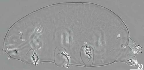
Figure 17. Drawing of Echiniscoides trichosus sp. nov. Scale bar in μm.
Figure 18. Detailed morphology of Echiniscoides trichosus sp. nov. (all but A in SEM): A. specimen in toto (DIC), B. dorsal sculpturing in the form of microtrichia, C. cephalic body portion with peribuccal cirri in frontal view, D. cephalic body portion with peribuccal cirri in lateral view, E. anterolateral body portion of paratypic female. Scale bars in μm.
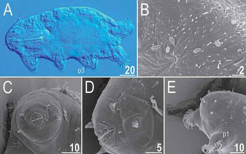
Figure 19. Dorsal sculpturing of Echiniscoides trichosus sp. nov. in close-up (SEM). Scale bars in μm.
Figure 20. Detailed morphology of Echiniscoides trichosus sp. nov. (all but D in SEM): A. specimen in toto in dorsal view, B–C. specimens in toto in lateroventral view, D. claws III and sense organ on leg III (PCM), E. claws III, F. claws IV. Scale bars in μm.
Figure 21. Habitus of Echiniscoides hispaniensis Kristensen & Hallas, Citation1980 stat. nov. (SEM): A. female in lateral view, B. female in anterolateral view, C. male in ventral view, D. female in ventral view. Scale bars in μm.
Figure 22. Detailed morphology of Echiniscoides hispaniensis (SEM): A. cephalic body portion with peribuccal cirri in lateral view, B. cephalic body portion with peribuccal cirri in frontal view, C. peribuccal cirri and secondary clava in close-up, D. cirrus A and primary clava, E. caudal body portion, F. cirrus E and dorsal sculpturing in close-up. Scale bars in μm.
Figure 23. Detailed morphology of Echiniscoides hispaniensis (SEM): A. claws I, B. claws III, C. male gonopore. Scale bars in μm.
Figure 24. Cuticular morphotypes within Echiniscoides (SEM): smooth cuticle – A–B. Echiniscoides sigismundi from Julebæk Strand (Sjælland, neotype locality), rugose cuticle – C. an undescribed Echiniscoides species from Los Angeles. Scale bars in μm.
Table IX. Main characteristics of described species of Echiniscoides.