Figures & data
Table I. Codes and origin of new sequences used in molecular study. For each specimen, the specimen catalogue number (GBIF code), the sample information (country, locality, coordinates and date of sampling), the GenBank accession number of the COI sequence, and nomenclature of genetic sequences (GenSeq; follows Chakrabarty et al. Citation2013) are provided.
Table II. Morphological characters of larvae that distinguish Centroptilum volodymyri sp. nov. from C. luteolum (Müller, 1776) and C. pirinense Ikonomov, Citation1962. Distinct differential characters are in bold.
Table III. Genetic distances (COI) between sequenced species of Centroptilum, Cloeon and Procloeon and within them, using Kimura two-parameter.
Figure 1. Larva of Centroptilum volodymyri sp. nov., Georgia, holotype. (a, b), Body in lateral view. Scale bars: 1 mm.
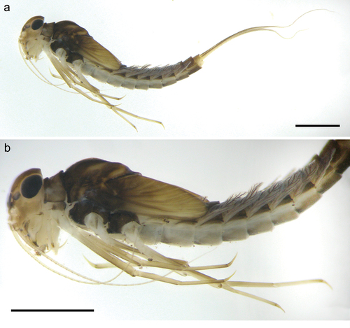
Figure 2. Larva of Centroptilum volodymyri sp. nov., Iran. (a) Body in dorsal view; (b) body in lateral view; (c) head and part of thorax in lateral view. Scale bars: 1 mm.
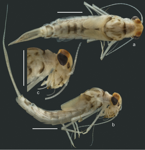
Figure 3. Larvae of Centroptilum volodymyri sp. nov., paratype (a, c, e) and C. luteolum (Müller, 1776) (b, d, f). (a, b) Scapus and pedicellus; (c) flagellum, basal part; (d) flagellum, central part; (e, f) surface of frons. Scale bars: a, c, d = 20 μm; b = 30 μm; e = 10 μm; f = 100 μm.
Figure 4. Larvae of Centroptilum volodymyri sp. nov., paratype (a–e) and C. luteolum (Müller, 1776) (f). (a) Labrum; (b) galea-lacinia; (c, d) mandibles, left (c) and right (d); (e, f) maxillary palp. Scale bars: a, b, e, f = 30 μm; c, d = 100 μm.
Figure 5. Larvae of Centroptilum volodymyri sp. nov., paratypes. (a, b) Labrum, ventral (a) and dorsal (b) view; (c) hypopharynx (left side – dorsal view; right side – ventral view); (d, e) glossa and paraglossa, dorsal (d) and ventral (e) view; (f) maxilla; (g, h) labial palp, ventral (g) and dorsal (h) view.
Figure 6. Larvae of Centroptilum volodymyri sp. nov., paratype (a, c, e) and C. luteolum (Müller, 1776) (b, d, f). (a, b) Pronotum, dorsal surface; (c, d) hind femur, dorsal surface; (e, f) hind tibia, dorsal surface. Scale bars: a–c, e, f = 30 μm; d = 10 μm.
Figure 7. Larvae of Centroptilum volodymyri sp. nov., paratype (a, c, e, g) and C. luteolum (Müller, 1776) (b, d, f). a, b = hind pretarsal claw (apex of claw not visible in (a)); (c, d) denticles of hind pretarsal claw; (e) surface of sternum IV; (f) surface and posterior margin of sternum VI; (g) surface and posterior margin of sternum VII. Scale bars: a = 50 μm; b = 100 μm; c–e, g = 30 μm; f = 10 μm.
Figure 8. Larvae of Centroptilum volodymyri sp. nov., paratypes. (a) Fore leg; (b) middle leg; (c) hind leg.
Figure 9. Larvae of Centroptilum volodymyri sp. nov., paratype (a, c, e, g) and C. luteolum (Müller, 1776) (b, d, f, h). Surfaces and posterior margins of terga: (a, b) tergum II; (c, d) tergum IV; (e, f) tergum VI; (g, h) tergum VIII. Scale bars: a, b = 20 μm; c–h = 30 μm.
Figure 10. Larvae of Centroptilum volodymyri sp. nov., paratypes. (a) Gill I; (b) gill II; (c) gill III; (d) gill IV; (e) gill V; (f) gill VI; (g) gill VII; (h) posterolateral part of tergum VII; (i) paraproct plate.
Figure 11. Larvae of Centroptilum volodymyri sp. nov., paratype (a, c, e) and C. luteolum (Müller, 1776) (b, d, f). (a, b) Apical part of paraproct; (c) surface of paraproct; (d) tergum X and caudal filaments; (e) caudal filaments, basal part; (f) caudal filament, medial part. Scale bars: a, b, e, f = 30 μm; c = 20 μm; d = 100 μm.
Figure 12. Eggs of Centroptilum volodymyri sp. nov., from paratype (a, c, e, g) and C. luteolum (Müller, 1776) (b, d, f, h); provided by Nicolás Ubero-Pascal). (a–d) General view of egg; (e, f) chorionic surface; (g, h) micropyle. Scale bars: a, c = 30 μm; b = 80 μm; d = 20 μm; e, g = 5 μm; f = 10 μm; h = 5 μm.
Figure 13. Habitats of Centroptilum volodymyri sp. nov. in Georgia, Iran and Turkey. (a, b) Small lentic waterbody, Georgia, Adjara, type locality (April 2016; photos by A. Martynov); (c, d) small brook, left tributary of Shalmanrud River, Guilan Province, Iran (May 2016; photos by J. Bojková); (e, f) small stream, Rize Province, Turkey (August 2012; photos by D. Palatov).
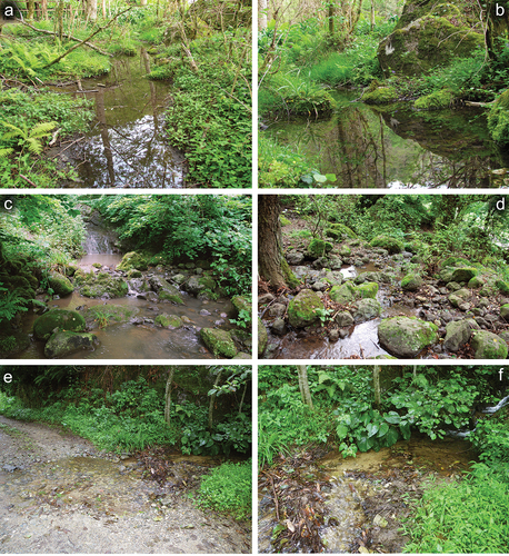
Figure 14. Map of Centroptilum volodymyri sp. nov. distribution. Red star – type locality; green pentagrams - non-type localities.
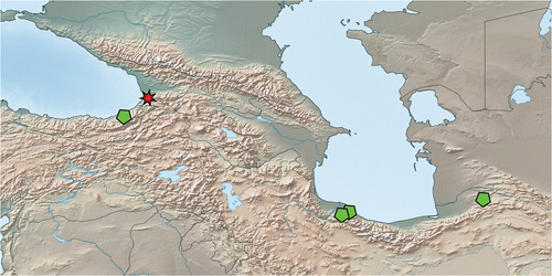
Figure 15. Maximum likelihood tree including several representatives of the genus Centroptilum. Bootstrap supports (BS) are indicated on branches for Centroptilum volodymyri sp. nov. and C. luteolum (Müller, 1776).