Figures & data
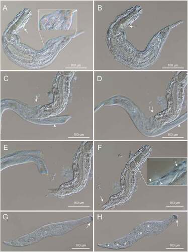
Figure 1. Differential interference contrast photomicrographs documenting the predation of Paraturbanella teissieri (Gastrotricha) by the dileptid Pseudomonilicaryon marinum (Ciliophora). (A, B) P. teissieri engulfed by P. marinum as seen at different focal planes; dotted lines trace the predator cytostome and the bilobed caudum of the engulfed prey; arrows indicate one of the two lateral adhesive organs of the prey; the insert highlights the predator “hairy” surface. (C, D) regurgitation of the prey; the predator cytostome (arrow) and its proboscis (arrowhead) are indicated. (E, F) the definitive separation between prey and predator; (E) P. teissieri appears completely immotile while P. marinum begins to regain its normal shape; (F) P. teissieri showing partially digested caudal lobes (arrow) and an uninjured anterior region as denoted (insert) by still well-shaped anterior adhesive tubes (arrow) and pristine tubes of the accessory adhesive organ (arrowhead). (G, H) the predator freed from the prey showing a fully functional cell body, in which the proboscis tip includes an agglomerate (arrow) of dark granules and the trunk accommodates some contractile and digestive vacuoles (asterisks). See text for further details.