Figures & data
Figure 1. SEM micrographs of Trogoderma granarium hastisetae: (A) hastiseta, lateral view; (B) tufts of hastisetae on the seventh and eighth abdominal tergites of the larva; (C) detail of the insertion of the hastiseta on the larval integument; (D) detail of a detached hastiseta showing the breaking point on the pedicel; (E) group of detached hastisetae illustrating how the rupture of the hastiseta occurs exclusively at the level of the pedicel; (F) detail of the rosettes that constitute the shaft of the hastiseta.
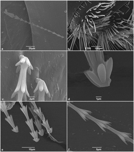
Figure 2. SEM micrographs of Trogoderma granarium hastisetae: (A) detail of the shaft between the ultimate rosette and the head of the hastiseta showing the set of irregular scales; (B) head of the hastiseta, frontal-lateral view; (C) detail of the longitudinal processes of the head of the hastiseta showing the knurls and the longitudinal depression (black arrows); (D) frontal view of the head the hastiseta illustrating the apical circular depression (black arrows).
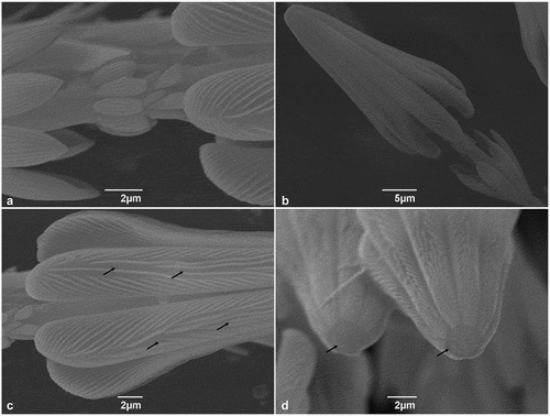
Figure 3. Ultrastructure of the larval tergite in transverse section; white rectangles – socket of the hastisetae, cu - cuticle, ep - epithelial cell, ne - neuron, tr- tracheoles.
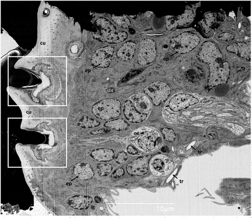
Figure 4. (A) Ultrastructure of the socket of the hastiseta in transverse section; (B) SEM micrograph of the cuticle of the point of insertion of the hastisetae on the larval tergite, taken from the endocuticle perspective. ht - hastiseta (pedicel), od - outer segment of sensory cell dendrite, m - microvilli, rls - receptor lymph space, tb - tubular body, tor - tormogen cell.
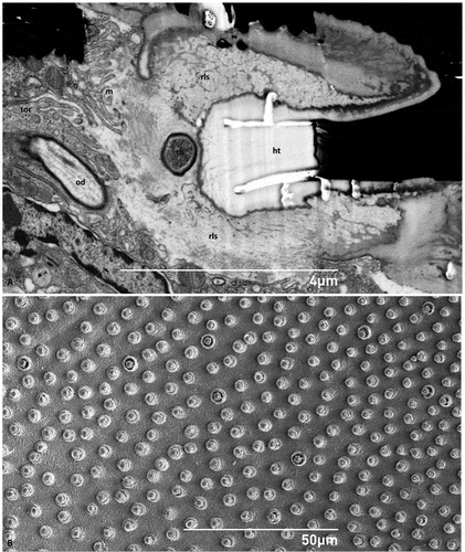
Figure 5. (A) Sagittal section of the socket of the hastiseta (pedicel missing) highlighting the lateral cuticle process probably involved in anchoring the socket; (B) detail of a convoluted branch of the outer dendrite; (C) sagittal section of the neurosensorial apparatus connected to the socket constituted by one neuron, its dendrite and the associated thecogen cell; (D) section showing the tubular bundle inside the tubular body; (E) detailed view of the connecting cilium. bb-basal body, cil - cilia, cr - ciliary rootlet, cs - ciliary sinus, ds - dendritic sheath, id - inner segment of sensory cell dendrite, lcp - lateral chitin process, ne - neuron, od - outer segment of sensory cell dendrite, tb - tubular body, the - thecogen cell, tub - tubular bundle.
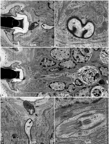
Figure 6. Ultrastructural features of neuronal and epithelial cells. (A-B) Sections of the neuron showing the rounded and central nucleus, mitochondria, Golgi and sparse rough endoplasmic reticulum; (C) detail of perikaryon shown in B illustrating a multivesicular body near a mitochondrion; (D-E) epithelial cells with irregularly shaped nucleus (note the abundance of mitochondria and the highly developed rough endoplasmic reticulum appearing as whorls); (F) close up of an elongated mitochondrion, and rough endoplasmic reticulum. go - Golgi, mt- mitochondrion, mvb- multivesicular body, nu - nucleus, rer - rough endoplasmic reticulum.
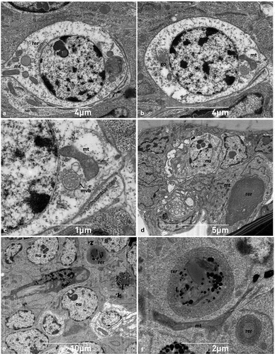
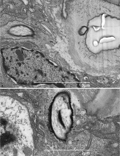
Figure 7. Scanning electron micrograph of the sagittal section of the socket of the hastiseta: A- B. Details of the apical microvillated membrane in hastisetae accessory glands. mv - microvilli, ve - vesicles.