Figures & data
Figure 1. Scanning electron microscopy (SEM) of apterous viviparous female of S. yushanensis general morphology: (a) habitus, (b) head and pronotum, (c) thorax, Pr-pronotum, Ms-mesonotum, Mt-metanotum.
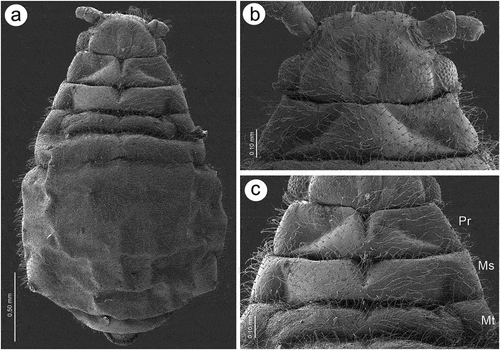
Figure 2. SEM of apterous viviparous female of S. yushanensis general morphology: (a) siphunculus densely covered by setae, (b) lateral side of the end of abdomen showing perianal structures with well-visible anal plate (ap), genital plate (gp) and poorly visible cauda (arrowhead), (c) back side of the end of abdomen with visible perianal structures and spiracles, VII-abdominal tergite VII, VIII-abdominal tergite VIII, c-cauda, ap-anal plate, arrow-rudimentary gonaphyses, gp-genital plate, (d) chaetotaxy of cauda and anal plate, (e) cauda and anal plate from the dorsal view, (f) ultrastructure of rudimentary gonapophyses.
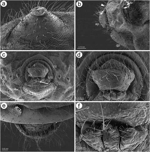
Figure 3. SEM of apterous viviparous female of S. yushanensis dorsal chaetotaxy and cuticle characters: (a) head chaetotaxy, (b) structure of the basal part of head trichoid sensilla and their sockets, (c) pronotum smooth cuticle surface, (d) surface of cuticle of the rest of thorax with numerous denticles, (e) ultrastructure of the cuticle surface and denticles, (f) dorsal abdominal chaetotaxy, (g, h) dorsal abdominal cuticle sculpture, (i) ultrastructure of the cuticle sculpture, (j) structure of the lateral abdomen cuticle and spiracles openings, (k) ultrastructure of the basal part of abdominal trichoid sensillum with the socket, (l) ultrastructure of the socket opening.
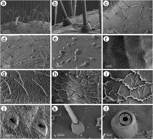
Figure 4. SEM of alate viviparous female and alatoid nymph of S. yushanensis: (a) dorsal side of alate viviparous female, (b) lateral side of alate viviparous female, (c) dorsal side of last instar alatoid nymph, (d) lateral side of last instar alatoid nymph.
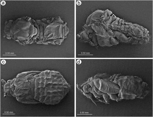
Figure 5. SEM of general morphological characters of alate viviparous female S. yushanensis: (a) head and pronotum, (b) dorsal side of head with large compound eye, lateral ocellus (star) and triommatidium (asterisk), (c) lateral side of head with large compound eye, lateral ocellus (star) and triommatidium (asterisk), (d) ultrastructure of triommatidium, (e) siphunculus on large sclerotic cone, (f) suphuncular opening with operculum, (g) lateral side of the end of abdomen showing siphunculus (s), abdominal segment VIII (VIII), short cauda (c), anal plate (ap) and large genital plate (gp), (h) waxy secretion on the body, (i) ultrastructure of the waxy secretion.
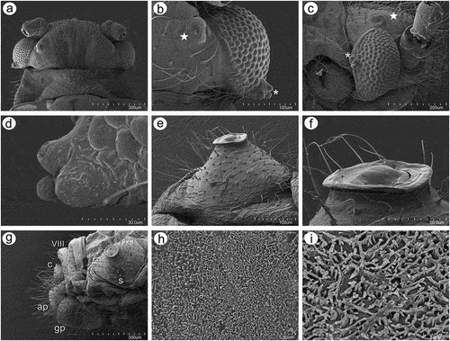
Figure 6. SEM of body surfaces of alate viviparous female of S. yushanensis: (a) pedicel with polygonal strengthenings, (b) ultrastructure of the strengthenings on the pedicel, (c) head cuticle with trichoid sensilla, (d) surface of abdominal dorsum, (e) ultrastructure of the dorsal cuticle, (f) structure of trichoid sensillum on the abdomen, (g) ultrastructure of the cuticle of ABD VII and VIII, (h) ultrastructure of trichoid sensilla socket and projections on the cuticle, (i) ultrastructure of dorsal cuticle with and without waxy secretion.
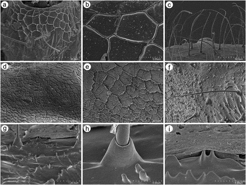
Figure 7. SEM of general morphological characters of alatoid nymph of S. yushanensis: (a) head, (b) dorsal side of pedicel with campaniform sensillum (star), (c) compound eye with larger dorsal edge and triommatidium, (d) dorsal area of eye cuticle with small openings (arrow), (e, f) ultrastructure of the possible ampulla sensilla, (g) trichoid sensilla of ANT III, (h) small multiporous placoid sensilla (secondary rhinaria) on ANT IV, (i) ultrastructure of the secondary rhinarium, (j) big multiporous placoid sensillum (primary rhinarium) on ANT V, (k) ultrastructure of porous membrane of the sensillum, (l) ANT VI sensilla, (m) ultrastructure of small multiporous placoid sensillum on ANT VI, (n) ultrastructure of type II trichoid sensilla on the tip of PT, (o) ultrastrusture of sunken coeloconic sensilla.
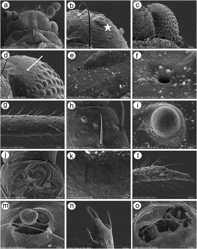
Figure 8. SEM of body surfaces and characters of alatoid nymph of S. yushanensis: (a) dorsal side of thorax with wing buds, (b) thoracic cuticle and trichoid sensilla, (c) dorso-lateral side of proximal part of fore wing bud with numerous openings in the cuticle, (d, e) ultrastructure of the openings in the cuticle, (f) lateral side of fore wing bud, (g) trichoid sensilla on the wing bud, (h) cuticle of the middle part of fore wing bud, (i, j) cuticle and trichoid sensilla of ABD I–III and ABD VII–VIII, (k, l) cuticle and trichoid sensilla of ABD IV–VII, (m) siphunculus, (n, o)perianal structures showing ABD VIII (VIII), short cauda (c), anal plate (ap) and genital plate bud (arrow).
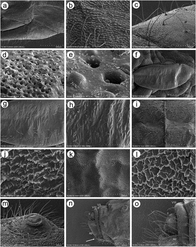
Figure 9. SEM of antennal sensilla of apterous viviparous female of S. yushanensis: (a) antennal flagellum general view, (b) ANT III with type I trichoid sensilla, (c) ANT IV with type I trichoid sensilla and one small multiporous placoid sensillum (Orange) – secondary rhinarium, (d) ANT V with type I trichoid sensilla, one small multiporous placoid sensillum (Orange) – secondary rhinarium and big multiporous placoid sensillum (yellow) primary rhinarium, (e) ANT VI with type I trichoid sensilla, type II trichoid sensilla (violet), big multiporous placoid sensillum (yellow) – major rhinarium, small multiporous placoid sensilla (green) and sunken coeloconic sensilla (pink) – accessory rhinaria.
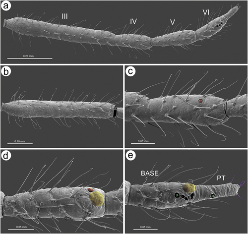
Figure 10. SEM of antennal trichoid sensilla of apterous viviparous female of S. yushanensis: (a) structure of type I trichoid sensilla with very fine often curved apices, (b) ultrastructure of type I trichoid sensillum and its socket, (c) ANT VI PT with small multiporous placoid sensillum and type II trichoid sensilla on the tip, (d) type II trichoid sensilla on the tip of ANT VI PT as apical (ap) and subapical (sap)setae, (e) structure of type II trichoid sensilla, (f) ultrastructure of type II trichoid sensilla apices.
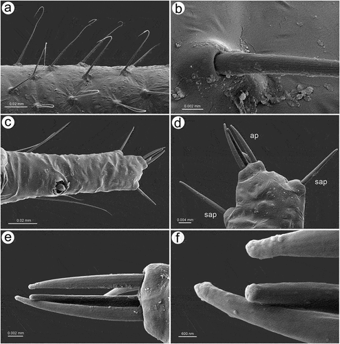
Figure 11. SEM of antennal placoid and coeloconic sensilla of apterous viviparous female of S. yushanensis: (a), ultrastructure of small multiporous placoid sensillum on ANT IV, (b) small and big multiporous placoid sensilla on ANT V, (c) ultrastructure of small multiporous placoid sensillum on ANT V, (d) ultrastructure membrane of small multiporous placoid sensillum on ANT V, (e) sensilla types on ANT VI BASE: type I trichoid sensilla (blue), big multiporous placoid sensillum (yellow) – major rhinarium, small multiporous placiod sensillum (green), two kinds of sunken coeloconic sensilla (pink) – accessory rhinaria, (f) group of two types of sunken coeloconic sensilla, (g) ultrastructure of small multiporous placoid sensillum on the BASE, (h) ultrastructure of small multiporous placoid sensillum on the PT, (i) type I sunken coeloconic sensillum (with short projections) and type II sunken coeloconic sensillum (with long projections).
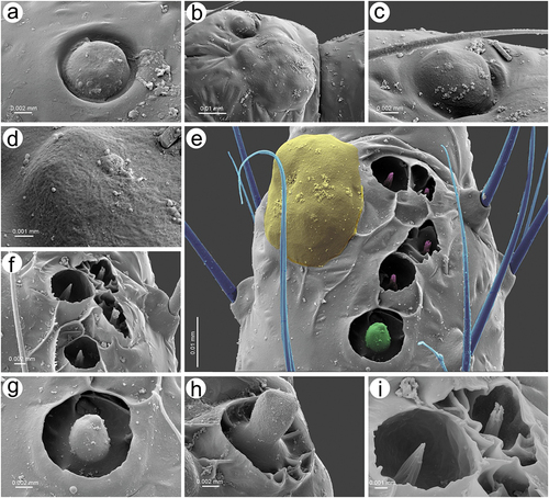
Figure 12. SEM of pedicel sensilla of alate viviparous female of S. yushanensis: (a) ventral side of pedicel with visible bare rhinariolum, (b) ultrastructure of the rhinariolum coeloconic-like sensillum with short projections, (c) dorso-lateral side of pedicel with campaniform sensillum (star) and a probable second type of sensillum on the edge (arrowhead), (d) ultrastructure of campaniform sensillum with small pore (asterisk), (e) edge of latero-dorsal side of pedicel with visible campaniform sensillum and the opening of a second type sensillum, (f) ultrastructure of the second type sensillum opening.
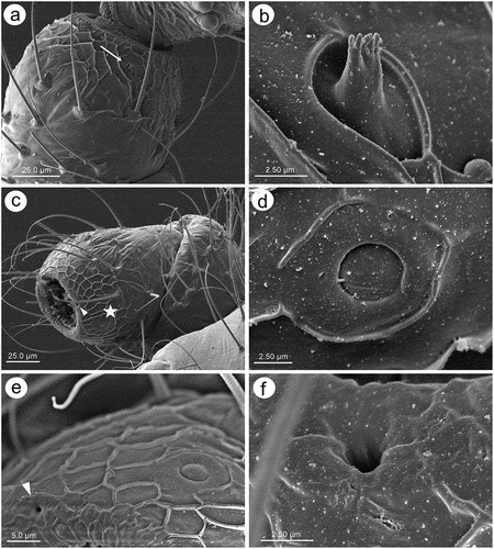
Figure 13. SEM of antennal flagellum sensilla of alate viviparous female of S. yushanensis: (a) fragment of ANT III with numerous small multiporous placoid sensilla (secondary rhinaria), (b, c) structure of secondary rhinaria and type I trichoid sensilla on ANT III, (d) different sizes of small multiporous placoid sensilla on ANT III with one of a size of big multiporous placoid sensillum (star), (e) small multiporous placoid sensilla n ANT IV od different sizes (star), (f) small multiporous placoid sensilla (secondary rhinaria) and big multiporous placoid sensilla (primary rhinarium) on ANT VI, (g, h) ultrastructure of small multiporous placoid sensillum with well visible sclerotic collar. Arrowhead showing the border between the cuticle and porous membrane, (i) ultrastructure of the porous membrane of small sensilla, (j) big multiporous sensillum with openings on the membrane (arrows), (k) structure of the sensillum membrane with the openings, (l) ultrastructure of porous membrane and the additional opening.
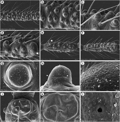
Figure 14. SEM of antennal flagellum sensilla of alate viviparous female of S. yushanensis: (a) ultrastructure of type I trichoid sensilla, (b) sensilla types on ANT VI: type I trichoid sensilla (unpigmented), small multiporous placoid sensilla (Orange) – secondary rhinaria, big multiporous placoid sensillum (major rhinarium), small multiporous placoid sensilla (green) – accessory rhinaria, sunken coeloconic sensilla (pink) – accessory rhinaria, type II trichoid sensilla (violet), (c) structure of type II trichoid sensilla, (d) topography of primary rhinaria on ANT VI. The largest is big multiporous placoid sensillum – major rhinarium (yellow), accessor rhinaria are divided into two general groups, sunken coeloconic sensilla in one group (pink) between two small multiporous placoid sensilla (green) in polar position, all are surrounded by type I trichoid sensilla (blue), (e) ultrastructure of small multiporous sensillum (accessory rhinarium), (f) ultrastructure of type I sunken coeloconic sensillum, (g) ultrastructure of type II coeloconic sensillum, (h) ultrastructure of type II trichoid sensilla, (i) ultrastructure of type II trichoid sensillum apex.
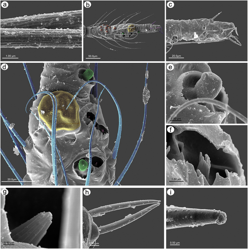
Figure 15. SEM of mouthparts sensilla of apterous viviparous female of S. yushanensis: (a) ventral side of ultimate rostral segments (URS) with numerous trochoid sensilla (arrow) on RIV and type III basiconic sensilla on the tip of RV (asterisk), (b) lateral side of URS with numerous trichoid sensilla (solid arrow), type II basiconic sensilla (dotted arrow) on RIV and type III basiconic sensilla on the tip of RV (asterisk), (c) dorsal side of URS with numerous trochoid sensilla (arrow) on RIV and type III basiconic sensilla on the tip of RV (asterisk), (d) structure of trichoid sensilla, (e) ultrastructure of basal part of trichoid sensillum and its socket, (f, g) ultrastructure of type II basiconic sensilla, (h) RV with type III basiconic sensilla and visible styletes (s), (i) four pairs of type III basiconic sensilla visible on the dorsal side of RV, (j) seven pairs of type III basiconic sensilla visible on the lateral side of RV, (k) six pairs of type III basiconic sensilla visible on the ventral side of RV, (l) ultrastructure of type III basiconic sensilla with molting pore on the basal part (star).
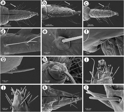
Figure 16. SEM of mouthparts characters and sensilla of alate viviparous female of S. yushanensis: (a) labium (rostrum) general view showing all five rostral segments, (b) trichoid sensilla on RII, (c) ultrastructure of socket and basal part of trichoid sensillum, (d) ultrastructure of middle part of trichoid sensillum, (e) ultrastructure of apical part of trichoid sensillum, (f) cuticle of the labial segment, (g) ventral side of ultimate rostral segments (URS) with numerous long trichoid sensilla and basiconic sensilla (asterisk), (h) proximal part of RIV with one pair of type II basiconic sensilla (arrows), (i) structure of type II basiconic sensillum, (j) ultrastructure of the socket of type II basiconic sensillum, (k) ultrastructure of the middle part of type II basiconic sensillum, (l) ultrastructure of the apical part of type II basiconic sensillum, (m) ultrastructure of sockets of trichoid sensilla, (n) RV with type III basiconis sensilla on the very apex, (o) dorso-lateral side of the apical part of RV with 7 visible pairs of basiconic sensilla, (p) ultrastructure of the basal part of type III basiconic sensillum with molting pore (star), (q) ultrastructure of apical part of basiconic sensillum with flat apex, (r) dorsal side of URS with numerous long trichoid sensilla and basiconic sensilla (asterisk), (s) lateral side of RV with type III basiconis sensilla on the very apex, (t), lateral side of the apical part of RV with 8 pairs of basiconic sensilla (u), ultrastructure of apical part of basiconic sensillum with rounded apex.
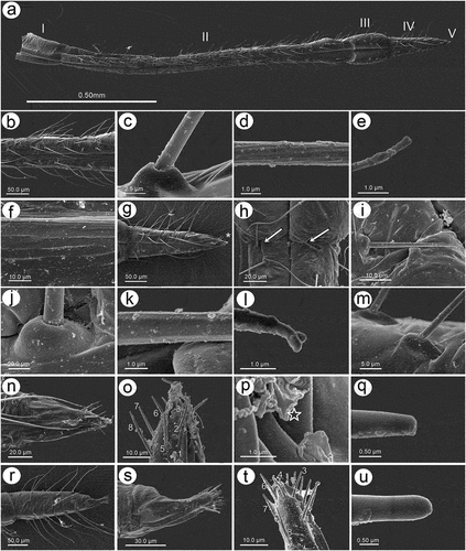
Figure 17. SEM of fore wings of alate viviparous female of S. yushanensis: (a) general view of the ventral side of fore wing with convex media (M1, M2), concave cubital veins (Cu1a, Cu1b) and visible claval fold (arrow), (b) general view of dorsal side of fore wing with concave media and convex cubital veins, (c) distal part of pterostigma and radial sector, (d, e) numerous campaniform sensilla near to the wing articulation, (f, g) ultrastructure of campaniform sensilla, (h) campaniform sensilla along inner side of Sc+R + M+ Cu (arrows), (i) scale-like elements on wing membrane on the inner surface, (j) ultrastructure of scale-like element, (k) scale like elements on the outer surface, (l) ruffled structures forming convex veins, (m) ultrastructure of the ruffled structures, (n) ultrastructure of scale-like element and membrane covered by wax secretion.
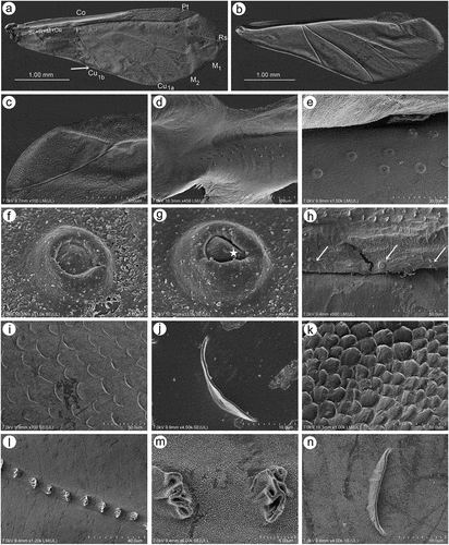
Figure 18. SEM of hind wings of alate viviparous female of S. yushanensis: (a) general view of the hind wing, (b) membrane and scale-like elements structure, (c) numerous small campaniform sensilla near the wing articulation (dotted rectangle), (d) ultrastructure of campaniform sensilla, (e) ultrastructure of the central part of campaniform sensilla with well-visible pore, (f) ventral side of claval area with five hamuli, (g) scale-like elements in the claval area, (h) dorsal side of claval area and hamuli.
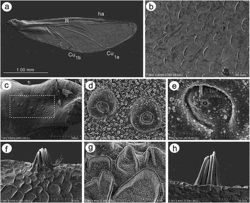
Figure 19. SEM of legs sensilla of apterous viviparous female of S. yushanensis: (a) campaniform sensilla on the inner side of hind trochanter (solid arrow) and hind femur (dotted arrow), (b) ultrastructure of campaniform sensillum on trochanter with visible pore (star), (c) trichoid sensilla on hind femur, (d) ultrastructure of tibial trichoid sensilla and their sockets, (e) trichoid sensilla on hind tibia, (f) ultrastructure of tibial trichoid sensillum and its socket, (g) hind tarsus, (h) two kinds of sensilla on ventral side of HTI – long sensilla (dotted arrow) and short, peg-like sensilla (solid arrow), (i) apical part of HT II with claws and residual, almost invisible parempodia (empodial setae) (arrow), (j) first segment of fore tarsus with largest number of peg-like sensilla, (k) first segment of middle tarsus with medium number of peg-like sensilla, (l) first segment of hind tarsus with smallest number of peg-like sensilla, HT I-first segment of hind tarsus, HT II-second segment of hind tarsus, cl-claws.
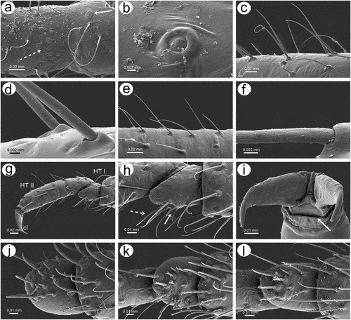
Figure 20. SEM of legs sensilla of alate viviparous female of S. yushanensis: (a) campaniform sensillum on hind trochanter, (b) ultrastructure of trochanteral campaniform sensillum with visible pore (star), (c) campaniform sensilla on hind femur, (d) ultrastructure of femoral campaniform sensillum with visible pore (star), (e, f) most probable ampulla sensillum (arrowhead) on the inner side of hind femora in the vicinity of campaniform sensilla, (g) ultrastructure of campaniform sensillum on the inner side of hind femora, (h) ultrastructure of probable ampulla sensillum, (i) trichoid sensilla on hind tibia, (j) ultrastructure of tibial trichoid sensilla, (k) trichoid sensilla on hind tibia, (l) hind tarsus, (m) first segment of hind tarsus with two kinds of sensilla on the ventral side, (n, o, p) apical part of HT II with very short almost residual parempodia (arrow) (empodial setae), (q) first segment of fore tarsus with largest number of peg-like sensilla, (r) first segment of middle tarsus with medium number of peg-like sensilla, (s) first segment of hind tarsus with smallest number of peg-like sensilla.
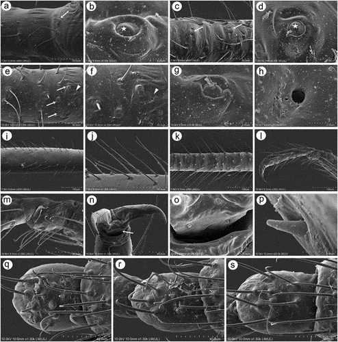
Figure 21. Morphological features of dorsal, ventral and lateral sides of alive specimens of Sinolachnus on the basis of S. yushanensis: (a) apterous viviparous females, (b) alate viviparous females, (c) alatoid nymph, (d) first instar larva.
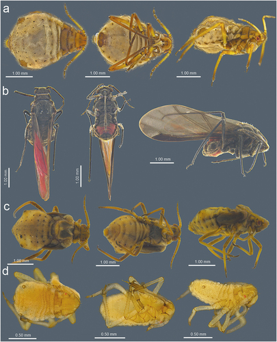
Table I. Measurements (in mm) of the examined apterous viviparous females of Sinolachnus.
Table II. Measurements (in mm) of known alate viviparous females of Sinolachnus.
Figure 22. Known apterous viviparous females of Sinolachnus: (a) S. rubi comb. nov., (b) S. yushanensis sp. nov.
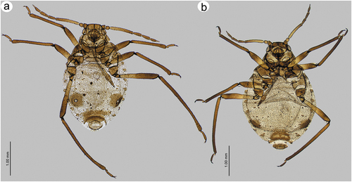
Figure 23. Alate viviparous females of Sinolachnus: (a) S. elaeagnensis, (b) S. nipponicus sp. nov., (c) S. niitakayamensis, (d) S. plurisensoriatus comb. nov., (e) S. takahashii sp. nov., (f) S. yushanensis sp. nov.
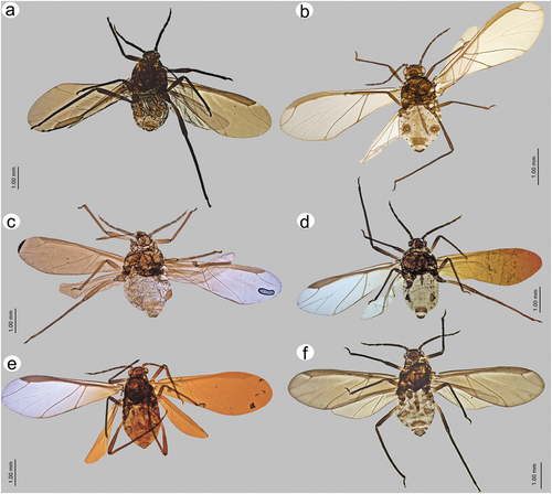
Figure 24. Morphological features of alate viviparous female of S. elaeagnensis: (a) antenna, (b) sensilla structure on ANT III, (c) ANT V with sensilla, (d) ANT VI with sensilla, (e) ultimate rostral segments, (f) hind wing sensilla, (g) dorsal abdominal cuticle, (h) first segment of fore tarsus, (i) first segment of middle tarsus, (j) first segment of hind tarsus, (k) dorsal abdominal chaetotaxy, (l) SIPH, (m) genital plate.
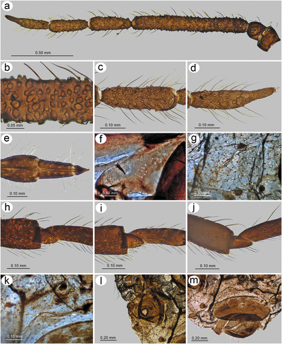
Figure 25. Morphological features of alate viviparous female of S. niitakayamensis: (a) antenna, (b) sensilla structure on ANT III, (c) ANT V with sensilla, (d) ANT VI with sensilla, (e) ultimate rostral segments, (f) hind wing sensilla, (g) dorsal abdominal cuticle, (h) first segment of fore tarsus, (i) first segment of middle tarsus, (j) first segment of hind tarsus, (k) dorsal abdominal chaetotaxy, (l) SIPH, (m) genital plate.
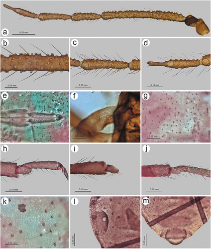
Figure 26. Morphological features of alate viviparous female of S. nipponicus sp. nov.: (a) antenna, (b) sensilla structure on ANT III, (c) ANT V with sensilla, (d) ANT VI with sensilla, (e) ultimate rostral segments, (f) hind wing sensilla, (g) dorsal abdominal cuticle, (h) first segment of fore tarsus, (i) first segment of middle tarsus, (j) first segment of hind tarsus, (k) dorsal abdominal chaetotaxy, (l) SIPH, (m) genital plate.
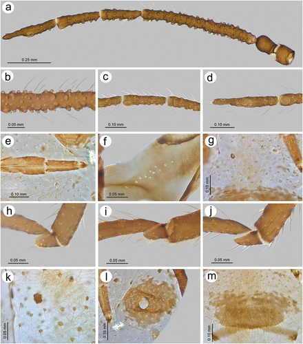
Figure 27. Morphological features of alate viviparous female of S. plurisensoriatus comb. nov.: (a) antenna, (b) sensilla structure on ANT III, (c) ANT V with sensilla, (d) ANT VI with sensilla, (e) ultimate rostral segments, (f) hind wing sensilla, (g) dorsal abdominal cuticle, (h) first segment of fore tarsus, (i) first segment of middle tarsus, (j) first segment of hind tarsus, (k) dorsal abdominal chaetotaxy, (l) SIPH, (m) genital plate.
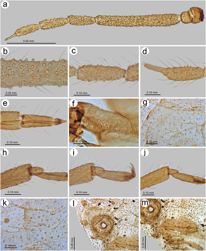
Figure 28. Morphological features of apterous viviparous female of S. rubi comb. nov.: (a) antenna, (b) sensilla on ANT IV, V and VI, (c) ANT VI with sensilla, (d) ultimate rostral segments, (e) mesosternal furca, (f) dorsal thoracic cuticle, (g) HT II, (h) first segment of fore tarsus, (i) first segment of middle tarsus, (j) first segment of hind tarsus showing the peg-like sensillum, (k) SIPH, (l) dorsal abdominal chaetotaxy, (m) dorsal abdominal cuticle.
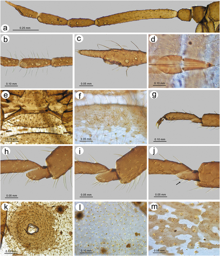
Figure 29. Morphological features of alate viviparous female of S. takahashii sp. nov.: (a) antenna, (b) sensilla structure on ANT III, (c) ANT V with sensilla, (d) ANT VI with sensilla, (e) ultimate rostral segments, (f) hind wing sensilla, (g) dorsal abdominal cuticle, (h) first segment of fore tarsus, (i) first segment of middle tarsus, (j) first segment of hind tarsus, (k) dorsal abdominal chaetotaxy, (l) SIPH, (m) genital plate.
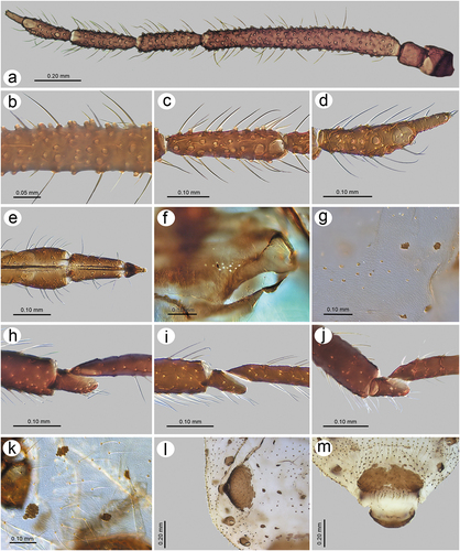
Figure 30. Morphological features of apterous viviparous female of S. yushanensis sp. nov.: (a) antenna, (b) sensilla on ANT IV, V and VI, (c) ANT VI with sensilla, (d) ultimate rostral segments, (e) mesosternal furca, (f) dorsal thoracic cuticle, (g) HT II, (h) first segment of fore tarsus, (i) first segment of middle tarsus, (j) first segment of hind tarsus showing the peg-like sensillum, (k) SIPH, (l) dorsal abdominal chaetotaxy, (m) dorsal abdominal cuticle.
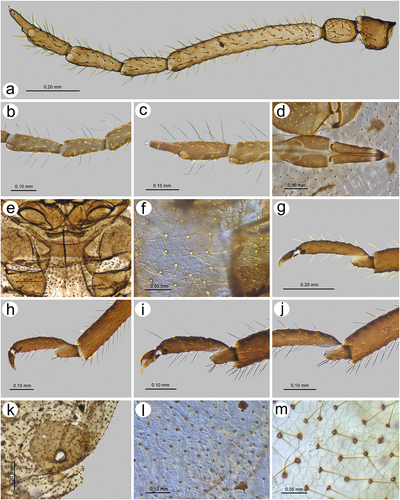
Figure 31. Morphological features of alate viviparous female of S. yushanensis sp. nov.: (a) antenna, (b) sensilla structure on ANT III, (c) ANT V with sensilla, (d) ANT VI with sensilla, (e) ultimate rostral segments, (f) hind wing sensilla, (g) dorsal abdominal cuticle, (h) first segment of fore tarsus, (i) first segment of middle tarsus, (j) first segment of hind tarsus, (k) dorsal abdominal chaetotaxy, (l) SIPH, (m) genital plate.
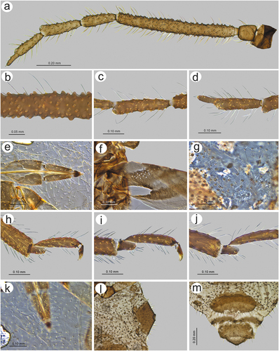
Figure 32. Biology of S. yushanensis: (a, b) colony of numerous apterous viviparous females and one alate viviparous female showing the pigmentation. Freshly molted individuals are characterized by shiny dorsum in contrast to the matt ones, (c, d) freshly molted apterous viviparous female and alate viviparous female visited by ants, (e) colony of mostly larvae of apterous viviparous females and nymphs of alate viviparous females and a freshly molted alate viviparous females with pale wings, (f) adult apterous viviparous female attacked by a neuropteran larva.
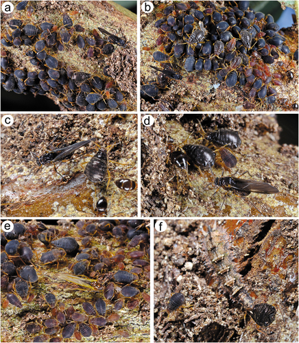
Figure 33. Fore wings comparison of alate viviparous females of Sinolachnus: (a) S. elaeagnensis, (b) S. niitakayamensis, (c) S. nipponicus sp. nov., (d) S. plurisensoriatus omb. nov., (e) S. takahashii sp. nov., (f) S. yushanensis sp. nov., (g) S. taiwanus (redrawn from Tao Citation1989).
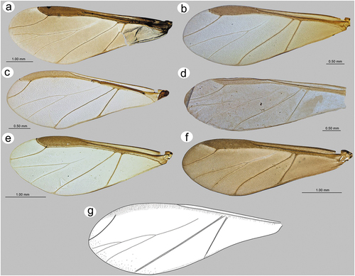
Figure 34. Comparison of Sinolachnus and remaining Tuberolachnini apterous viviparous females: (a) Sinolachnus yushanensis, (b) Nippolachnus piri, (c) Pyrolachnus pyri, (d) Tuberolachnus salignus.
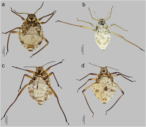
Figure 35. Comparison of Sinolachnus and remaining Tuberolachnini alate viviparous females: (a) Sinolachnus yushanensis, (b) Nippolachnus piri, (c) Pyrolachnus pyri, (d) Tuberolachnus salignus.
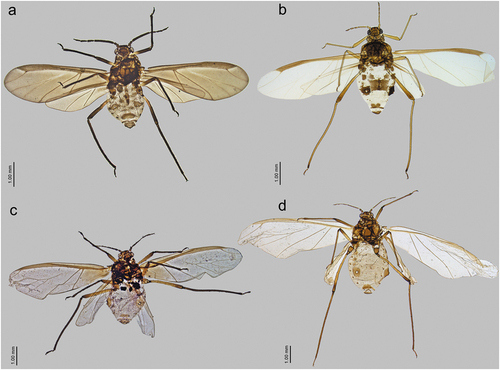
Figure 36. Comparison of Sinolachnus and remaining Tuberolachnini antennal features: (a) ANT of S. niitakayamensis, (b) ANT of N. piri, (c) ANT of P. pyri, (d) ANT of T. salignus, (e) ANT VI of S. niitakayamensis, (f) ANT VI of N. piri, (g) ANT VI of P. pyri, (h) ANT VI of T. salignus.
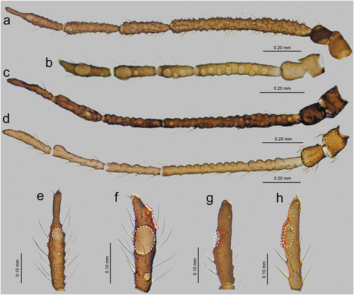
Figure 37. Comparison of Sinolachnus and remaining Tuberolachnini first segment of tarsi chaetotaxy and sense pegs: (a) FT I, (b) MT I, (c) HT I of S. niitakayamensis; (d) FT I, (e) MT I, (f) HT I of N. piri; (g) FT I, (h) MT I, (i) HT I of P. pyri; (j) FT I, (k) MT I, (l) HT I of T. salignus.
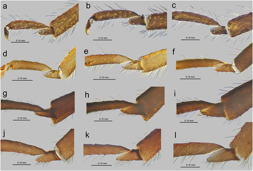
Figure 38. Comparison of Sinolachnus and Eotrama: (a) apterous viviparous female of E. moerickei, (b) alate viviparous female of E. moerickei, (c) apterous viviparous female of S. yushanensis, (d) alate viviparous female of S. yushanensis.
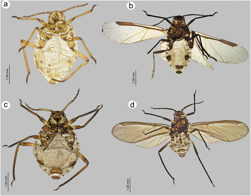
Figure 39. Comparison of Sinolachnus and Tramini: (a) antenna of Sinolachnus nipponicus, (b) antenna of Protrama radicis, (c) antenna of Eotrama moerickei, (d) ANT VI of S. nipponicus, (e) ANT VI of P. radicis, (f) E. moerickei, (g) first segment of fore tarsus of S. yushanensis, (h) first segment of fore tarsus of E. moerickei, (i) first segment of middle tarsus of E. moerickei, (j) first segment of fore tarsus of P. radicis, (k) first segment of middle tarsus of P. radicis, (l) first segment of fore tarsus of Trama troglodytes.
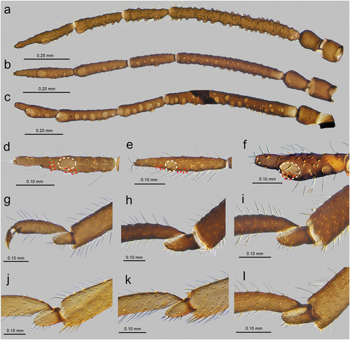
Figure 40. Distributional map of the species of Sinolachnus.