Figures & data
Table I. Species described in the Myxicola genus (*sensu Hayward & Ryland Citation1990) in the World Register of Marine Species (WoRMS).
Figure 1. Distribution of the genus Myxicola worldwide. For codes, see .
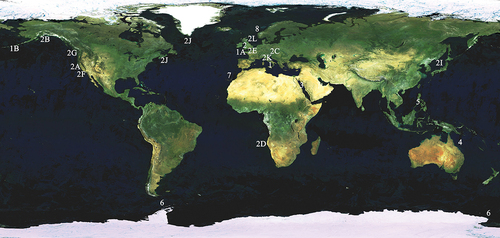
Figure 2. Myxicola sp. 1. (a) entire worm; (b) peristomial mid-lateral incision; (c, d) scheme of the peristomal ring: (c) ventral view; (d) dorsal view; (e) complex of ventral and dorsal lips; (f) radiolar tip; (g) scheme of radiolar section. Scale bars: b, f = 1 mm.
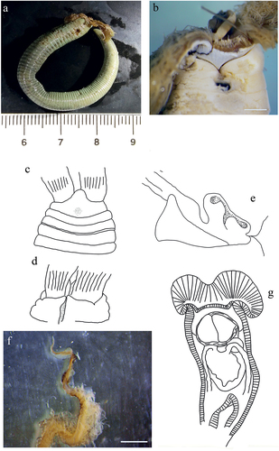
Figure 3. Myxicola sp. 1. (a) 1st setiger thoracic chaeta; (b) 4th setiger thoracic chaeta; (c) 24th abdominal chaeta; (d) 4th thoracic uncini; (e) 24th abdominal uncinus, lateral view and scheme of teeth above the main fang. Scale bar: 0.05 mm.
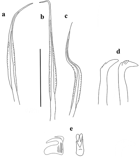
Figure 4. Myxicola sp. 2. (a) entire worm from Naples; (b, c) scheme of the peristomal ring: (b) ventral view; (c) lateral view; (d) complex of ventral and dorsal lips; (e) pygidium; (f) radiolar tip; (g) scheme of radiolar section. Scale bar: b, c = 2 mm; d, f = 1 mm, e = 0.5 mm.
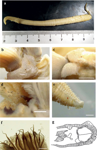
Figure 5. Myxicola sp. 2. (a) 1st setiger thoracic chaeta; (b) 4th setiger thoracic chaeta; (c) 24th abdominal chaeta; (d) 4th thoracic uncinus; (e) 24th abdominal uncini, lateral and front view. Scale bar: 0.05 mm.
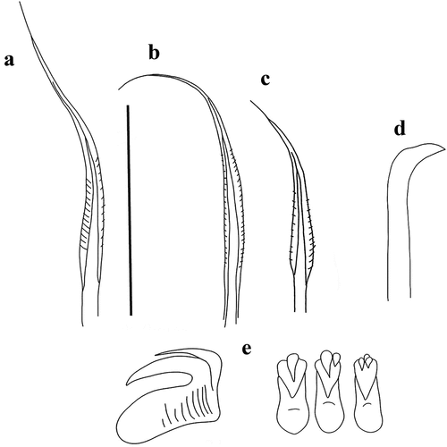
Figure 6. Myxicola cosentinii. (a) entire worm; (b, c) peristomial ring, ventral and lateral view; (d) complex of ventral and dorsal lips; (e) pygidium; (f) radiolar tip; (g) radiolar eyes. Scale bars: b, c = 2 mm; d, f = 1 mm; e = 0.5 mm; g = 0.1 mm.
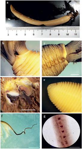
Figure 7. Myxicola cosentinii. (a) scheme of radiolar section; (b) 1st setiger thoracic chaeta; (c) 4th setiger thoracic chaeta; (d) 24th abdominal chaeta; (e) 4th setiger thoracic uncinus; (f) 24th abdominal uncini, lateral and front view; Scale bar: b–f = 0.05 mm.
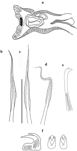
Figure 8. Myxicola giuliae. (a) entire worm; (b, c) peristomial ring, ventral and lateral view; (d) complex of ventral and dorsal lips; (e) pygidium; (f) radiolar tip; (g) scheme of radiolar section. Scale bars: b, c, f = 2 mm; d = 1 mm; e = 0.5 mm.
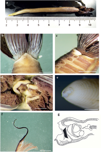
Figure 9. Myxicola giuliae. (a) 1st setiger thoracic chaeta; (b) 4th setiger thoracic chaeta; (c) 24th abdominal chaeta; (d) 4th setiger thoracic uncinus; (e) 24th abdominal uncini, lateral and front view. Scale bar: 0.05 mm.
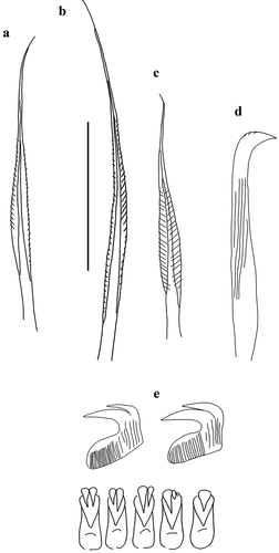
Figure 10. Myxicola cataldoi. (a) entire worm; (b, c) peristomial ring, ventral and lateral view; (d) complex of ventral and dorsal lips; (e) pygidium; (f) radiolar tip; (g) scheme of radiolar section. Scale bars: b, d, f = 1 mm; c = 2 mm; e = 0.5 mm.
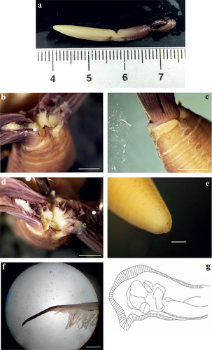
Figure 11. Myxicola cataldoi. (a) 1st setiger thoracic chaeta; (b) 4th setiger thoracic chaeta; (c). 24th abdominal chaeta; (d) 4th setiger thoracic uncinus; (e) 24th abdominal uncini, lateral and front view. Scale bar: 0.05 mm.
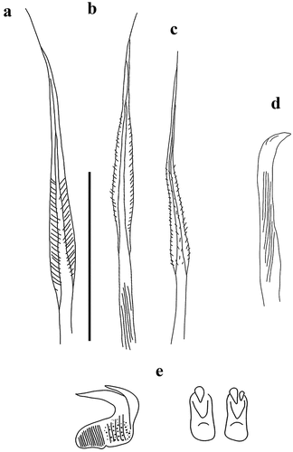
Figure 12. Myxicola mikacae. (a) entire worm; (b–d) peristomial ring: (b) ventral, (c) dorsal and (d) lateral view; (e) complex of ventral and dorsal lips; (f) photo of peristomial ring; (g) radiolar tip; (h) pygidium; (i) scheme of radiolar section. Scale bars: f = 2 mm; g = 1 mm; h = 0.5 mm.
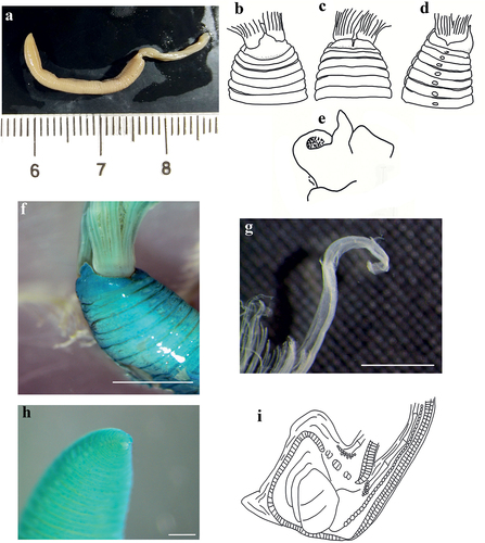
Figure 13. Myxicola mikacae. (a) 1st setiger thoracic chaeta; (b) 4th setiger thoracic chaeta; (c) 24th abdominal chaeta; (d) 4th setiger thoracic uncini; (e) 24th abdominal uncini, lateral and front view. Scale bar: 0.05 mm.
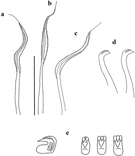
Figure 14. Myxicola pacifica. (a) entire worm; (b, c) peristomial ring, ventral and lateral view; (d) complex of ventral and dorsal lips; (e) radiolar tips; (f) scheme of radiolar section; (g) pygidium. Scale bars: b, c, e = 1 mm; g = 0.5 mm.
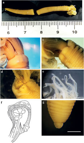
Figure 15. Myxicola pacifica. (a) 1st setiger thoracic chaeta; (b) 4th setiger thoracic chaeta; (c) 24th abdominal chaeta.; (d) 4th setiger thoracic uncinus of the original material: syntype MCZ ANNb-1879; (e) 4th setiger thoracic uncinus; (f) 24th abdominal uncinus of the original material: syntype MCZ ANNb-1879; G 24th abdominal uncini, lateral and front view; Scale bar: 0.05 mm.
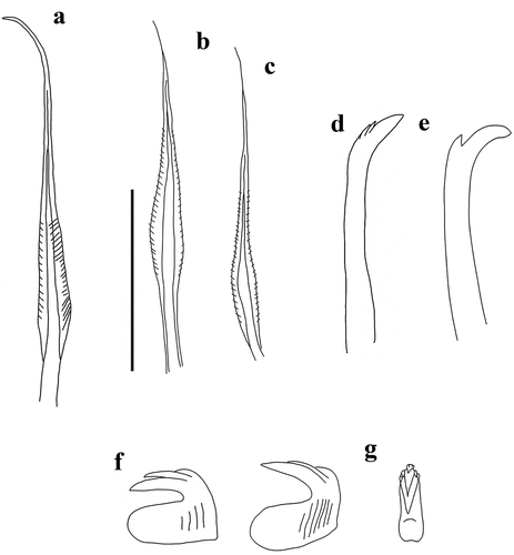
Table II. Distribution of character in the up to now recognized species of Myxicola genus.
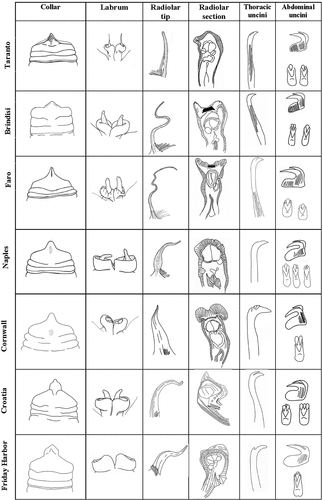
Figure 16. Comparative table of the main morphological characters of the Myxicola species studied.