Figures & data
Figure 1. Scolelepis tridentata (Southern, Citation1914), syntype (NHMUK 1914.12.12.29): A. Anterior end, dorsal view; palps, left notopodial postchaetal lamellae of chaetiger 3, right parapodia of chaetigers 1 and 2, and right notopodial postchaetal lamellae of chaetigers 3 and 4 missing. B. Anterior end, lateral view; C. Notopodial limbate capillary chaetae, chaetiger 35. D. Neuropodial hook of chaetiger 34. E. Ventral inferior capillary chaeta, chaetiger 35. F. Alternating capillary chaeta, chaetiger 35. Scale bars: A, B = 0.5 mm; C = 0.1 mm; D–F = 50 μm.
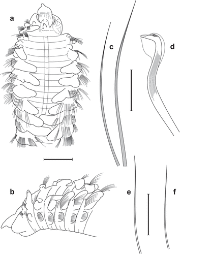
Figure 2. Scolelepis tridentata (Southern, Citation1914), syntypes (A–D – NMINH 1914.325.7; E – NMINH 1910.42.4): A. Parapodium of chaetiger 1, anterior view. B. Parapodium of chaetiger 2, anterior view. C. Parapodium of chaetiger 8, anterior view. D. Parapodium of chaetiger 21, anterior view. E. Parapodium of posterior chaetiger, anterior view. Scale bars: A–E = 0.2 mm.
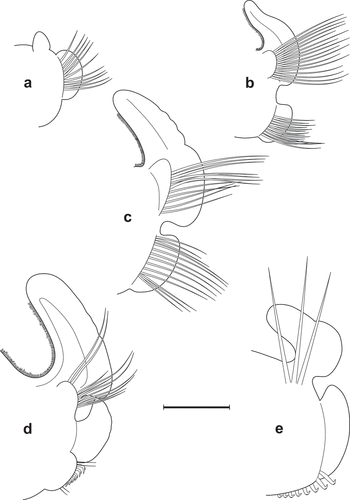
Figure 3. Scolelepis tridentata (Southern, Citation1914), syntype (NMINH 1914.325.7), LM micrographs, stained with MB: A. Pygidium, postero-latero-dorsal view; B, chaetigers 22–27, dorsal view. Scale bars: A = 0.25 mm, B = 0.2 mm.
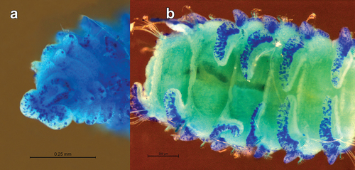
Figure 4. Scolelepis tridentata (Southern, Citation1914), syntype (NMINH 1914.325.7), SEM micrographs: A, Neuropodial hooks with hoods, lateral view; B, Neuropodial hook with three apical teeth, latero-apical view, hood removed; C, Neuropodial hooks with two and three apical teeth, latero-apical view, hoods removed; D, Neuropodial hook with two apical teeth, apical view, hood removed. Scale bars: A = 20 μm; B–D = 10 μm.
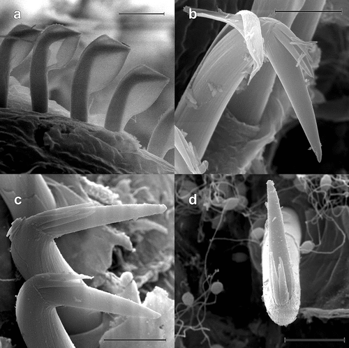
Figure 5. Scolelepis bellani n. sp., paratype (A–C, F – MGAB PLY0169), holotype (D – MGAB PLY0166), and voucher specimen (E – VS), LM micrographs, stained with MB: A. Anterior end, dorsal view, palps missing. B. Anterior end, lateral view, palps missing. C. Anterior end, frontal-dorsal view, palps missing. D. Middle body chaetigers (chaetigers 36–41), dorsal view, showing notopodial postchaetal lamellae divided into a superior flag-like process and inferior narrow part. E. Anterior end, lateral view, showing palps with papillate basal sheaths. F. Pygidium, dorsal view. Scale bars: A–D, F = 0.5 mm; E = 0.25 mm.
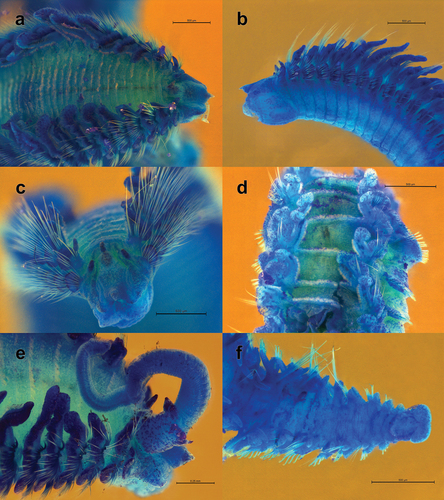
Figure 6. Scolelepis bellani n. sp., paratype (MGAB PLY0169): A. Parapodium of chaetiger 1, anterior view. B. Parapodium of chaetiger 2, anterior view. C. Parapodium of chaetiger 3, anterior view. D. Parapodium of chaetiger 14, anterior view. E. Parapodium of chaetiger 30, anterior view. Scale bars: A–E = 0.2 mm.
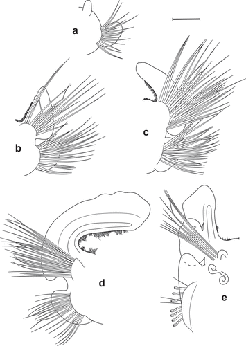
Figure 7. Scolelepis bellani n. sp., paratype (MGAB PLY0169): A. Parapodium of chaetiger 44, anterior view. B. Parapodium of posterior chaetiger, anterior view. C. Mid-part of a notopodial limbate capillary chaeta, chaetiger 30. D. Neuropodial hook of chaetiger 30. Scale bars: A, B = 0.2 mm; C, D = 20 μm.
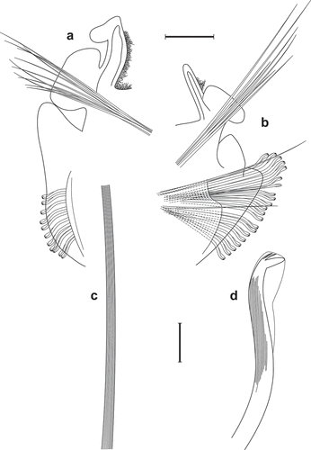
Figure 8. Scolelepis bellani n. sp., paratype (MGAB PLY0168), SEM micrographs: A. Anterior end, dorsal view, palps missing. B. Neuropodial capillary chaetae of chaetiger 4. C. Neuropodial hooded hooks of chaetiger 28, left latero-ventral view (dorsal side is to the left and the anterior end is down). D, Neuropodial hook with three apical teeth, latero-apical view, hood removed. E, Neuropodial hook with four apical teeth, frontal view, hood removed. F, Neuropodial hook with four apical teeth, latero-apical view, hood removed. Scale bars: A = 0.5 mm; B, C = 50 μm; D–F = 5 μm.
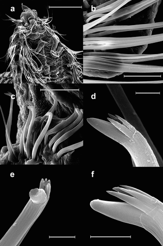
Figure 9. Results of the morphometric multivariate analysis of individuals belonging to six species of Scolelepis: A. Non-metric multidimensional scaling (nMDS) plot. B. Principal Component Analysis (PCA) plot. S. tridentata (green upward-pointing triangles), S. papillosa (blue circles), S. korsuni (blue downward-pointing triangles), S. quinquedentata (pink squares), S. bellani n. sp. (red diamonds), and S. cantabra (grey asterisks). Holotypes represented by hollow symbols.
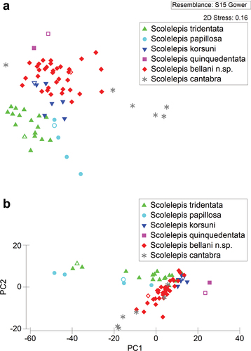
Table I. Results of ANOSIM pairwise comparisons between analysed species of Scolelepis, showing R statistics and p values.
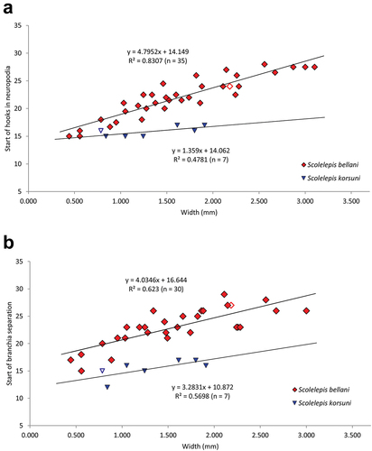
Figure 10. Scatter plots of relationships between: A. The body width (W) and the number of the first chaetiger bearing neuropodial hooded hooks (VH). B. The body width (W) and the number of the first chaetiger on which notopodial postchaetal lamella splits from branchia (Br) (red diamonds – Scolelepis bellani n. sp.; blue downward-pointing triangles – Scolelepis korsuni). Holotypes represented by hollow symbols.