Figures & data
Table I. GenBank codes of COI sequences produced in this work.
Table II. COI sequences of Heleomyzidae downloaded from BOLD and included in the analyses.
Figure 1. Heleomyza serrata adult (male). Lateral view (a), magnification of male genitalia (red box 1a)(b), head chaetotaxy in frontal view (c), head chaetotaxy in dorsal view (d), thorax chaetotaxy in dorsal view (e), prosternum (f), thorax chaetotaxy in lateral view (g) and magnification of the katepisternum (red box 1g)(h). Observation performed using a Keyence VHX-S90BE digital microscope (scale bar 500 µm).
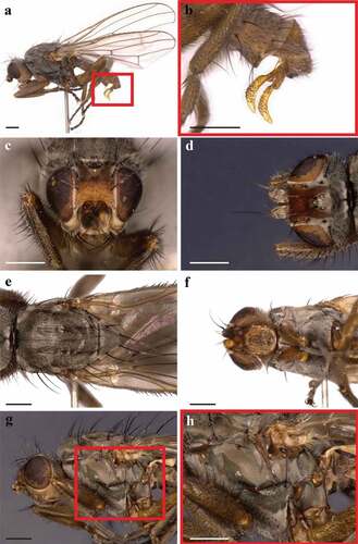
Figure 2. Heleomyza serrata puparium. Ventral (a), dorsal (b) and lateral (c) views. Observation performed using a Keyence VHX-S90BE digital microscope (scale bar 500 μm).
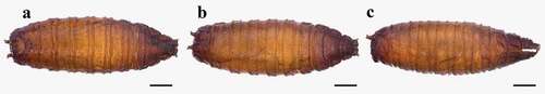
Figure 3. Heleomyza serrata puparium details. Anal plate (a), posterior spiracles (b,c), anterior spiracle (d) and intersegmental spicules (e). Observation performed using a Keyence VHX-S90BE digital microscope (scale bar 500 μm).
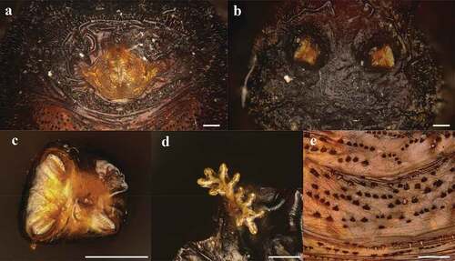
Figure 4. Heleomyza serrata puparium details. Anal plate (a), intersegmental spicules (b), posterior spiracle (c) and anterior spiracle (d). Observation performed using a SEM (scale bar is reported in each frame).
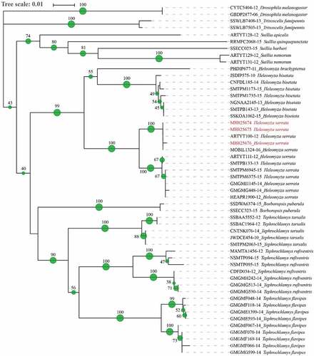
Figure 5. Phylogenetic tree of Heleomyzidae family. Neighbour joining method analysis of 563 bp sequence of the COI gene. The green spots and the number at each node indicate the bootstrap support. Sequences from this study are reported in red.