Figures & data
Table I. Primers with their original references used for sequencing of three molecular markers of Mesobiotus mandalori sp. nov.
Table II. Linear regression of morphometric traits in relation to buccal tube length in Mesobiotus mandalori sp. nov. SSIPm represents Stylet support insertion point (µm), BTEWm represents Buccal Tube External width (µm), BTIWm represents Buccal Tube Internal Width (µm), VLLm represents Ventral Lamina Length (µm), M1m represents Macroplacoid 1 length (µm), M2m represents Macroplacoid 2 length (µm), M3m represents Macroplacoid 3 length (µm), Mim represents Microplacoid length (µm), MRm represents Macroplacoid Row length (µm) and PRm represents Placoid Row length (µm). The simple linear regression is performed using Graphpad Prisn 8.0.
Table III. Linear regression of morphometric traits in relation to buccal tube length in Mesobiotus mandalori sp. nov. SSIPpt represents Stylet support insertion point (pt value), BTEWpt represents Buccal Tube External width (pt value), BTIWpt represents Buccal Tube Internal Width (pt value), VLLpt represents Ventral Laptina Length (pt value), PT1pt represents Macroplacoid 1 length (pt value), PT2pt represents Macroplacoid 2 length (pt value), PT3pt represents Macroplacoid 3 length (pt value), Mipt represents Microplacoid length (pt value), MRpt represents Macroplacoid Row length (pt value) and PRpt represents Placoid Row length (pt value). The simple linear regression is performed using Graphpad Prisn 8.0.
Figure 1. X–Y plots of raw data for SSIP (a); BTEW (b), VLL (c) and BTIW (d).
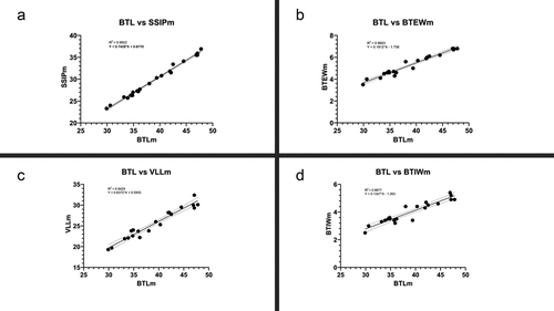
Figure 2. X–Y plots of pt value BTEW (a), SSIP (b), BTIW (c) and VLL (d).
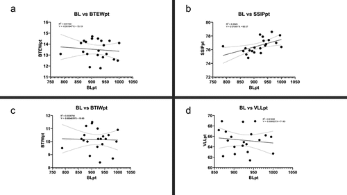
Table IV. Measurements [in µm] and pt values of selected morphological structures of Mesobiotus. mandalori sp. nov. Table contains measurements [in µm] and pt values of selected morphological structures of the newly described species. “N” stands for the number of specimens/structures measured; “RANGE” refers to measurements taken for the smallest and the largest structure among all measured specimens; SD stands for standard deviation.
Figure 3. Mesobiotus mandalori sp. nov. — Phase-contrast microscope (PCM) and scanning electron microscope (SEM images of habitus morphology: (a) — PCM dorso-ventral projection (holotype); (b) — SEM dorsal projection (paratype). Scale bars in μm.
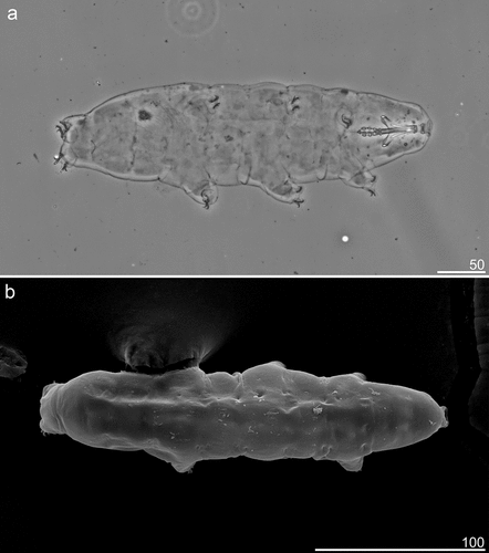
Figure 4. Mesobiotus mandalori sp. nov. —PCM images of claws I, III and IV: (a) — Claw I with smooth lunules (paratype); (b) —Claw III with smooth lunules (paratype); (c) — Claw IV with dented lunules (holotype). (d) — Lunules under claws IV with dented lunules (paratype). Filled flat arrowhead indicates lunules; filled indented arrowheads indicate the granulation; empty indented arrowheads indicate the indicates horseshoe structure connecting the anterior and the posterior claws; empty arrow indicates a single continuous cuticular bar under the claws; filled arrow indicates paired muscle attachments Scale bars in μm.
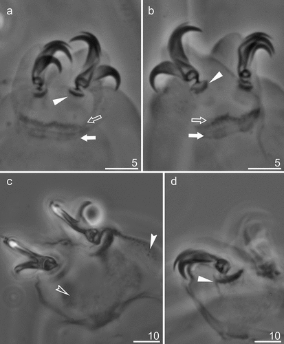
Figure 5. Mesobiotus mandalori sp. nov. —PCM images of the entire buccal apparatus: (a) — dorsal view of entire buccal apparatus (holotype); (b) — ventral view of entire buccal apparatus (holotype). Scale bars in μm.
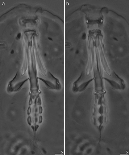
Figure 6. Mesobiotus mandalori sp. nov. —PCM images of oral cavity armature (OCA) and placoid morphology: (a) — dorsal view of OCA (paratype); (b) — ventral view of OCA (paratype); (c) — dorsal view of placoid morphology; (d) — ventral view of placoid morphology. Filled flat arrowheads indicate peribuccal lamellae; empty flat arrowheads indicate the first band of teeth; filled indented arrowheads indicate the second band of teeth; filled indented arrowheads indicate the third band of teeth, filled arrows indicate the middle ridge dorsal and ventral respectively; empty arrows indicate subterminal constrictions in the third macroplacoid. Scale bars in μm.
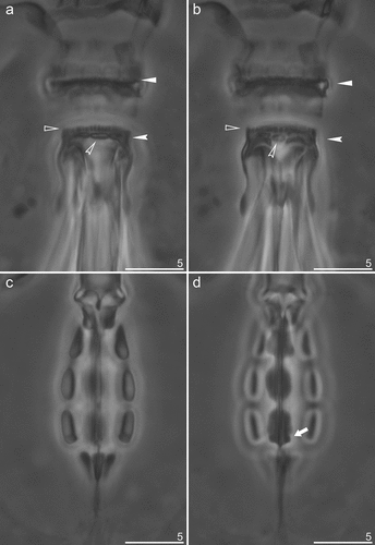
Table V. Measurements [in µm] of selected morphological structures of eggs of Mesobiotus mandalori sp. nov. Table contains measurements [in µm] structures of eggs of newly described species. Where “N” stands for the number of specimens/structures measured; “RANGE” refers to measurements taken for the smallest and the largest structure among all measured specimens; SD stands for standard deviation.
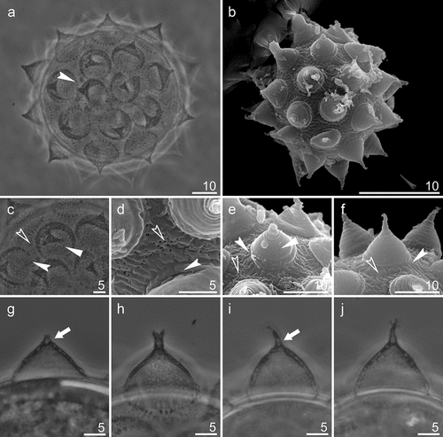
Figure 7. Mesobiotus mandalori sp. nov. —PCM and SEM images of the eggs, egg surface, egg processes surface and midsections: (a) PCM image of the eggs; (b) SEM image of the egg; (c) PCM image of the egg surface; (d) SEM image of the egg surface; (e-f) SEM images of the egg processes surface; (g-j) PCM images of the egg processes midsections. Filled flat arrowhead indicates reticular design; filled indented arrowheads indicate crowns of strong thickenings around the process base; empty indented arrowheads indicate pores;filled arrow indicates bubble-like internal structures, Filled flat arrowheads indicate reticular design; filled arrows crowns of strong thickenings around the process base; empty arrows indicate pores. Scale bars in μm.