
Figures & data
Figure 1. The habitats where parasitized spiders were collected: (a) ombrotrophic mire Młyńskie Bagno of the Izera Valley (SW Poland), (b) wet grassland near Kruszyniany (NE Poland). Photo by K. Wiśniewski (a) and U. Jabłońska (b).
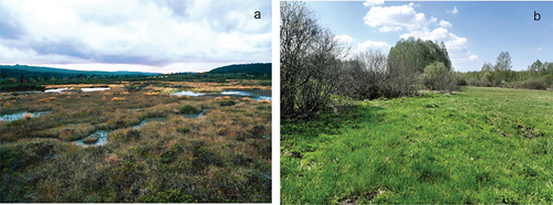
Table I. Characteristics of localities in which the parasitized spiders were collected, and sampling methods used.
Table II. Studied spider species and mermithid haplotypes (H1, H2), with sampling dates (for Izera Valley and Turczyn – pitfall trap exposure; in the case of Kruszyniany – sampling date and, in brackets, date of spider death). Number of specimens is indicated if more than one were observed. ns – sequence of 18S rDNA gene was not obtained, jv – juvenile, ♀ - female, ♂ - male.
Figure 2. Mermithids in: (a) Trochosa sp., (b, c) Alopecosa pulverulenta, (d, f) Pardosa paludicola, (e) Piratula hygrophila; (d, e) mermithids emergence from opisthosoma, (f) mermithid presence in pedicel. Photo by A. Kostro-Ambroziak.
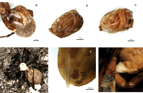
Figure 3. Epigyne (ventral view) of Piratula hygrophila in: (a) female parasitized by a mermithid, (b) unparasitized female. Photo by U. Jabłońska.

Figure 4. Epigyne (ventral view) of Pardosa paludicola in: (a, b) females parasitized by a mermithid, (c) unparasitized female. Photo by U. Jabłońska.
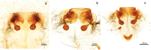
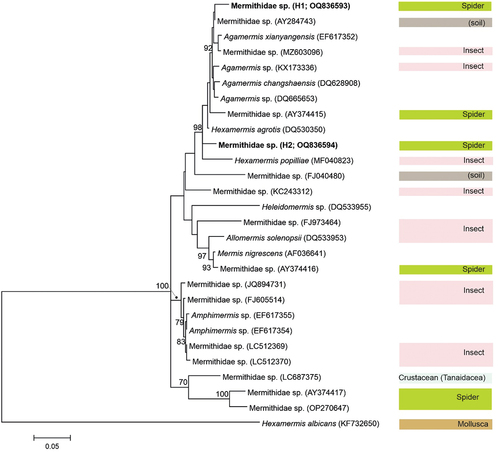
Figure 5. Maximum‐likelihood (ML) phylogenetic tree of Mermithidae based on 18S rDNA, including the new haplotypes obtained in the present study (marked in bold). Information on the known host group is presented in the right-hand column. Bootstrap values (≥70%) are indicated at the nodes. Scale bar indicates branch length in substitutions per site.
Supplemental Material
Download MS Word (24.9 KB)Data availability statement
The datasets generated during the current study are deposited in GenBank under accession number OQ836593 and OQ836594, and included in this published article and its supplementary information file.