Figures & data
Table 1. Body weights (g), blood glucose (mg/dl) and systolic BP (mm/Hg), locomotor activity, passive avoidance acquisition test phases and MWMT of the time spent in the target quadrant on day 5 of rats in 4- (4 M), 18-(18 M), 24-month-old (24 M) and agmatine-treated age-matching groups.
Figure 1. Passive avoidance retention test phases of young (4-month-old), middle-aged (18-month-old), aged (24-month-old) and agmatine (40 mg/kg)-treated matching groups (n = 12 rats in each group). Data were presented as mean ± SEM and *P < .05 and ***P < .001 compared to the young control group (4-month-old).
Figure 2. Escape latency (s) in MWMT results of 1–4 days by rats in young (4-month-old), middle-aged (18-month-old), aged (24-month-old) and agmatine (40 mg/kg)-treated matching groups (n = 12 rats in each group). Data were presented as mean ± SEM and **P < .01 and ***P < .001 compared to the young control group (4-month-old).
Figure 3. Representative image illustrating BDNF expression (arrows) in hippocampal formation. Control (a), 24-month (b) and 24-month + agmatine (c) groups in the CA1 region, and control (d), 24-month (e) and 24-month + agmatine (f) groups in the CA3 region. control (g), 24 month (h) and 24-month + agmatine (i) groups in the amygdala. BDNF expression was decreased in 24 month in the CA1 and CA3 regions of the hippocampus, and amygdala, whereas in 24-month + agmatine rats BDNF expression was similar to those in the control group in the all regions. Scale bars: 50 μm.
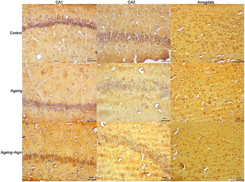
Table 2. Semi-quantitative distribution of BDNF-immunoreactive neurons of the CA1 and CA3 regions of the hippocampus and amygdala and eNOSimmunoreactivity of aorta in rats.
Table 3. Em (% of 10−6 M phenylephrine) values for carbachol, sodium nitroprusside and papaverine, Em values (g) for 80 mM KCl and pD2 values (–log EC50) for carbachol, and sodium nitroprusside in rings of thoracic aorta obtained from 4- (4 M), 18-(18 M), 24-month-old (24 M) and agmatine-treated age-matching group rats.
Figure 4. Carbachol concentration–response curves in isolated thoracic aortic rings pre-contracted with phenylephrine (10−6 M). The concentration–response curve for carbachol was shifted to the right with significantly lower values of Em and pD2 in the 24-month-old group than in 4-month-old controls (*P < .05). Impairment of relaxation in the agmatine treatment group was returned to that seen in the controls. Each point is expressed as a percentage of the contraction induced by phenylephrine and is represented as the mean ± SEM. The values in parentheses indicate the number of preparations used; *P < .05, different from the response of tissue rings from 4-month-old young control rats.
Figure 5. Sodium nitroprusside concentration–response curves in isolated thoracic aortic rings pre-contracted with phenylephrine (10−6 M). The relaxation response to SNP was similar among vascular tissues from all groups. Each point is expressed as a percentage of the contraction induced by phenylephrine and is expressed as the mean ± SEM. The values in parentheses indicate the number of preparations used.
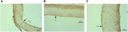
Figure 6. Representative light microscopy of control, 24-month-old and agmatine-treated 24-month-old groups in the endothelium of the aorta. Decreased eNOS (arrows) (b) immunoreactivity in the aorta in 24-month-old rats compared to 4-month-old controls (a) and increased eNOS (c) immunoreactivity in endothelial tissue in the agmatine-treated 24-month-old group compared to 24-month-old groups (b). Scale bars: 20 μm.