Figures & data
Figure 1. Schematic subcategorization of patients with different myositis-specific autoantibodies (MSAs). Because IIM patients with different MSAs have common clinical characteristics, each MSA-positive patient can be determined to be a subset of IIM. The horizontal axis shows the spectrum of skin disease and muscle involvement, and the vertical axis shows the acute or chronic type of ILD.
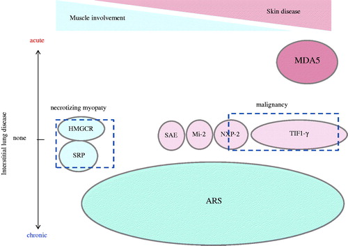
Table 1. The frequency and significance of myositis-specific autoantibodies in adult IIM patients.
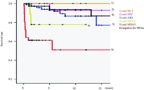
Figure 2. The survival rate of patient groups with different myositis-specific autoantibodies (MSAs). Overall, 245 Japanese IIM patients who visited our department were divided into groups, myositis-specific autoantibody-positive subsets (anti-Mi-2, anti-SRP, anti-ARS, anti-TIF1-γ, anti-MDA5 and negative for MSAs), and their survival rate was plotted.