
Figures & data
Figure 1. Representative photograph of healthy control.
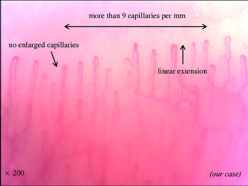
Figure 2. Representative photograph of irregularly enlarged capillary, giant capillary, and microhemorrhage.
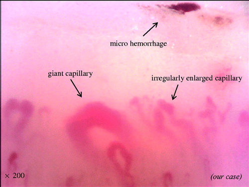
Figure 3. Representative photograph of disorganization of loss of capillaries, microvascular array, and capillary ramifications.
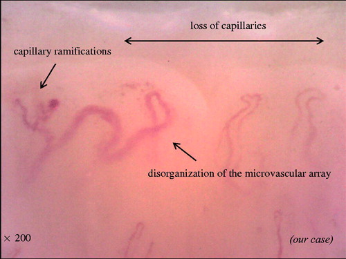
Figure 4. Representative photograph of ‘early pattern’.
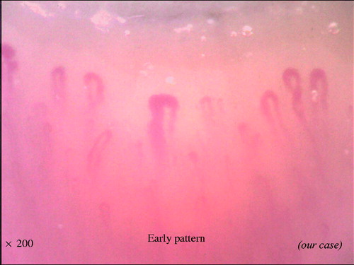
Figure 5. Representative photograph of ‘active pattern’.
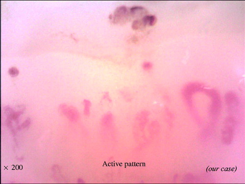
Figure 6. Representative photograph of ‘late pattern’.
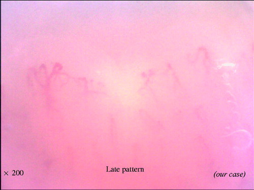
Figure 7. Comparison between NVC scleroderma spectrum abnormality and Steinbrocker radiographic stage score.
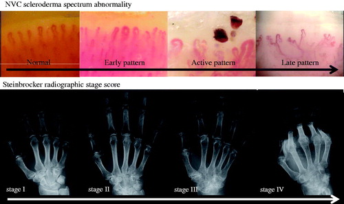
Table 1. Definition of each pattern of NVC scleroderma spectrum abnormalities.