
Figures & data
Table 1. Chemical compositions, manufacturers, and product names of the various materials used in this study.
Figure 1. Proximal view of the virtual model for endocrown restoration.
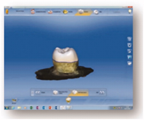
Figure 2. Cross sectional view of virtual model for endocrown restoration.
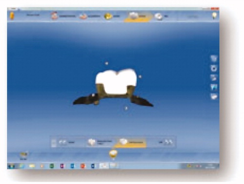
Figure 3. Ceramill COMP steromicroscope (90X magnification) A: before aging; B: after aging.
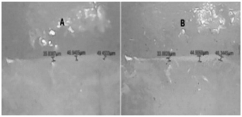
Figure 4. Cerasmart steromicroscope (90X magnification) A: before aging; B: after aging.
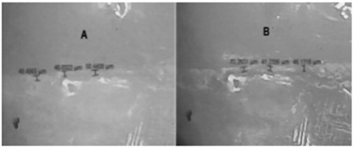
Figure 5. Mean marginal gap before and after thermo-mechanical aging via different bonding protocols for each material approach.
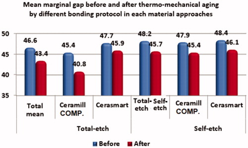
Table 2. Comparisons of the marginal gaps for the different material groups and bonding protocols.
Figure 6. Mean load required to fracture.

Table 3. Comparisons of the load required to fracture the endocrown with respect to the material used and different bonding protocols.
Data availability
All data supporting the reported results are available in a report available from the corresponding author upon request.