
Figures & data
Figure 1. Schematics of (B): two initial bacterial adhesion counting methods.
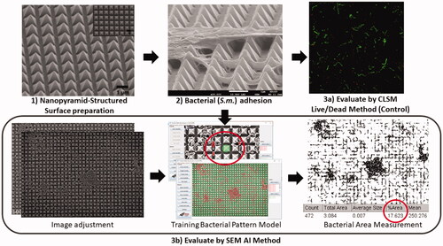
Figure 2. Demonstration of manually label the bacteria and background.
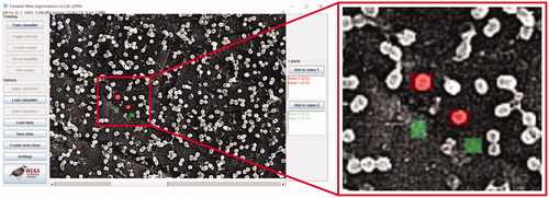
Figure 3. The area percentage of P.g. and F.n. occupied area after 1 h, 7 h and 24 h’ inoculation.
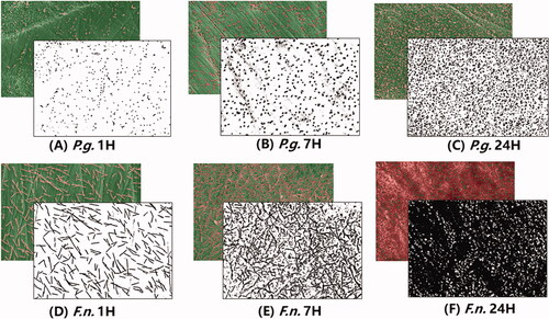
Figure 4. Area percentages of two bacteria in different time points and linear regression of the data.
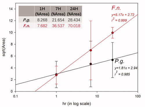
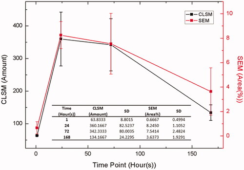
Figure 5. Summary of the bacterial adhesion results evaluated by CLSM (live plus died) and SEM images in different cell incubation times.
Supplemental material