
Figures & data
Figure 1. Experimental set-up using (a) the commercial SS-OCT (Santec IVS-300, Japan) and (b) the non-commercial SD-OCT (Queen Mary University of London, London, UK).
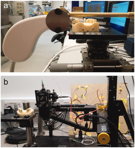
Figure 2. Details of measurement method: black lines cervically on the ceramic crown placed on the abutment tooth indicate the area for OCT imaging (left). B-scans (center) were obtained from five sites at the facial surface, 250 µm apart. On the B-scans, measurement sites were defined. Lines delimiting the marginal horizontal (a) and vertical (b) discrepancies were used to calculate the absolute marginal discrepancy (c), and measurement areas were delimited for assessment of internal fit. The internal cement gap was defined as the distance between two peaks on the gray value intensity plot (right).
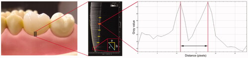
Figure 3. Box-plots representing (a) absolute marginal discrepancy (µm) and (b) internal cement gap (µm) of lithium disilicate crowns assessed by SD-OCT and SS-OCT.
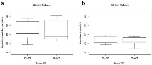
Figure 4. Box-plots representing (a) absolute marginal discrepancy (µm) and (b) internal cement gap (µm) of zirconia crowns assessed by SD-OCT and SS-OCT.
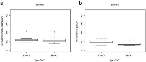
Figure 5. Percentage of measured and missing internal cement gap values for lithium disilicate and zirconia crowns according both OCT system.
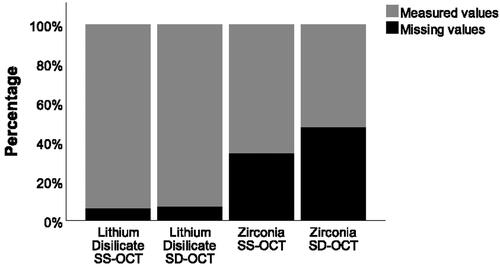
Table 1. Median and interquartile ranges (IQR) of height of marginal gap (i.e. straight vertical line from preparation margin towards crown margin) and overextended margins (i.e. straight horizontal line from preparation margin towards facial surface of the crown) of lithium disilicate and zirconia crowns using Swept Source OCT (SS-OCT) and Spectral Domain OCT (SD-OCT).