
Figures & data
Figure 1. Macro photos, scanning electron microscopy images and 3D surface topography of the three different test surfaces. Surface roughness (Sa) of the test surfaces was calculated from the obtained profilometry data, Ti: 0.25 ± 0.01 µm; TiF: 1.94 ± 0.16 µm; TCP: 9.34 ± 0.21 nm (n = 3).
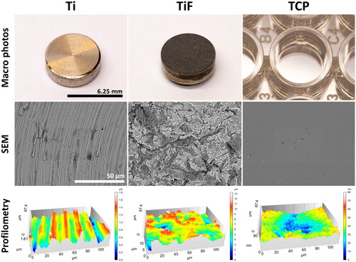
Figure 2. The number of irradiated human osteoblasts (hOBs) cultured in growth medium (GM) or differentiation medium (DM) on minimally rough machined titanium (Ti) and moderately rough fluoride-modified titanium (TiF), and the relative confluence of hOBs on tissue culture polystyrene (TCP), 21 days post-irradiation. Data represent mean values ± standard error of the mean (SEM) from three different experiments (n = 9) and are presented as % of unirradiated controls (red line) on the same surfaces (Ti, TiF or TCP) and in the same type of culture medium (GM or DM). The significance level is set to p values ≤0.05 toward control (red line) (a), toward 2 Gy (b) and toward 6 Gy (c).
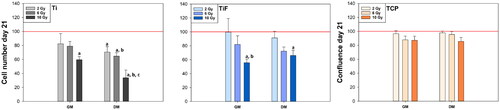
Figure 3. Confocal images from one of the experiments. Morphology of cell nuclei, collagen and actin production of hOBs cultured for 21 days in growth medium (GM) (n = 3) or differentiation medium (DM) (n = 3) on minimally rough machined titanium (Ti) and moderately rough fluoride-modified titanium (TiF), after no irradiation and after 10 Gy (A). Quantification of collagen (B) and actin (C) production of hOBs cultured for 21 days in GM (n = 3) or DM (n = 3), on Ti and TiF, after single doses of 2, 6 and 10 Gy. The data represent mean values ± standard error of the mean (SEM) from three different experiments and are presented as % of unirradiated controls (red line) on the same surfaces (Ti or TiF) and in the same type of culture medium (GM or DM). The significance level is set to p values ≤ 0.05 toward control (a), and toward 6 Gy (c).
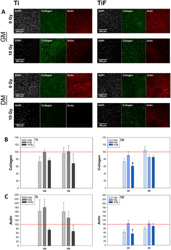
Figure 4. Alkaline phosphatase (ALP) activity measured in growth medium (GM) (n = 9) and differentiation medium (DM) (n = 9) of human osteoblasts (hOBs) on days 1 and 21 post-irradiation (A), and the effects of radiation on the mineral deposition of hOBs after 21 days of culture (B) on minimally rough machined titanium (Ti) (n = 6), moderately rough fluoride-modified titanium (TiF) (n = 6), and tissue culture polystyrene (TCP) (n = 9), after single doses of 2, 6 and 10 Gy. The data represent mean values ± standard error of the mean (SEM) from three different experiments and are presented as % of unirradiated controls (red line) on the same surface (Ti, TiF or TCP) and timepoint. The significance level is set to p values ≤ 0.05 toward control (a), toward 2 Gy (b) and toward 6 Gy (c).
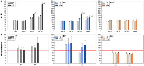
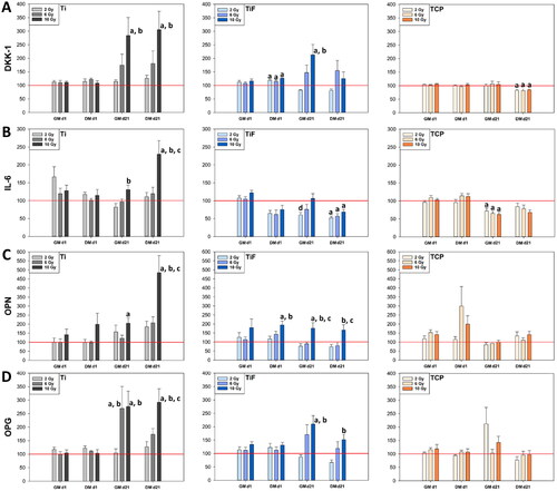
Figure 5. Secretion of osteogenic biomarkers; dickkopf-related protein (DKK-1) (A), interleukin-6 (IL-6) (B), osteopontin (OPN) (C) and osteoprotegerin (OPG) (D) to the cell culture medium (GM and DM) measured on days 1 and 21 post-irradiation, from human osteoblasts (hOBs) cultured on minimally rough machined titanium (Ti) (n = 9), moderately rough fluoride-modified titanium (TiF) (n = 9) and tissue culture polystyrene (TCP) (n = 9) after single doses of 2, 6 or 10 Gy. The data are presented as % of unirradiated controls (red line) on the same surfaces (Ti, TiF or TCP) and timepoints. Data were adjusted to the relative cell number on day 21 and represent mean values ± standard error of the mean (SEM) from three different experiments. The significance level is set to p values ≤ 0.05 toward control (a), toward 2 Gy (b), toward 6 Gy (c) and toward 10 Gy (d).
Supplemental Material
Download MS Word (442.4 KB)Data availability statement
The datasets generated and/or analyzed during the current study are available from the corresponding author on reasonable request.