Figures & data
Figure 1. Serial changes of the body weight during experiment. The ovariectomized (OVX) rats showed more rapid weight gain than sham-operated (Sham) rats. The LT4 treatment has no effect on change of body weight.
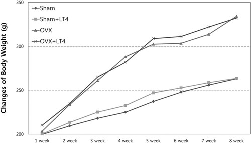
Table 1. Median values for DXA outcomes and comparisons among the experimental groups.
Figure 2. The difference in cross-sectional area (CSA), Feret’s diameter (FD) and area of the interstitium. (A) CSA were significantly higher in ovariectomized (OVX) groups than in Sham-operated (Sham) only group. (B) FD were significantly elevated in OVX groups compared with Sham groups regardless of LT4 treatment. (C) The areas of the interstitium were increasing with OVX and LT4 treatment. OVX with LT treatment showed most widened interstitial space. *, P < 0.05 compared with the Sham only operation group; ^, P < 0.05, compared with Sham with LT4 treatment; **, P < 0.05 compared with OVX only group.
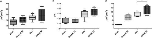
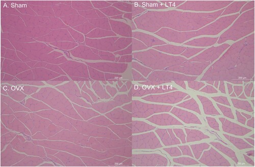
Figure 3. The representative hematoxylin-eosin staining of coronary section of soleus muscle in light microscope images of hematoxylin-eosin stained section of soleus muscle. Sham operated (Sham) only rats (A) showed most intact and dense morphology of the muscle (A). Sham with LT4 treatment (B) or OVX OVX without LT4 treatment (C) showed disorganized and muscle fibers and widening change of the interstitium compared with Sham group (A). OVX + LT4 rats (D) showed most significant decreased muscle volume, sparsely distributed muscle fibers and widened interstitium of soleus muscle.