
Figures & data
Figure 1. hAD-MSCs and cardiomyocyte purity identification. (A) hAD-MSCs cells; (B–F) Flow cytometry detection of CD44, CD73, CD105, CD90, CD34, CD45, CD11B, CD19 and HLA-DR expression in isolated cultured hAD-MSCs; (G) Cardiomyocytes; (H) The expression of a-actin in isolated and cultured cardiomyocytes was detected by flow cytometry; (I) Bar chart of statistical results.
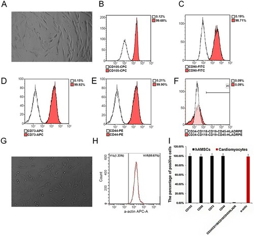
Figure 2. TIPE2 overexpressing adenoviral vector construction and infection. (A) Viral expression vector backbone; (B) Adenovirus packaging; (C) Transfection efficiency of adenovirus. (D) Empty virus and TIPE2 overexpressing virus respectively infect hAD-MSCs. (E) Detection of the TIPE2 expression by Western blot.
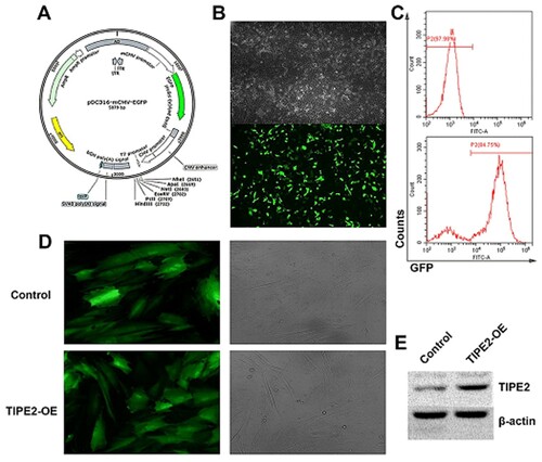
Figure 3. Cell viability assay. (A–D), Cell cycle assay; (E–H), Groups of cells. Apoptosis detection; (I) Statistical results of cell cycle; (J) Statistical results of apoptosis; (K) MTT results. The four groups of cells were (1) Contact co-culture: TIPE2 gene-modified hAD-MSCs. (2) Contact co-culture of null-hAD-MSCs. (3) Non-contact co-culture of TIPE2 gene-modified hAD-MSCs. (4) Non-contact co-culture of null-hAD-MSCs.
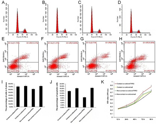
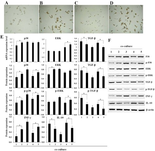
Figure 4. Detection of related gene and protein expression of immune responses. (A–D), vWF detection by immunohistochemistry, the four groups of cells were contact co-culture/TIPE2 (A), contact co-culture/null (B), non-contact co-culture/TIPE2 (C), and non-contact co-culture of/null (D). E, qRT-PCR and western blot detect the expression of p38, ERK, TGF-β, INF-γ, and IL-10. F, Western blot detect the protein expression. (1) contact co-culture/TIPE2; (2) contact co-culture/null; (3) cardiomyocytes; (4) non-contact co-culture/TIPE2; (5) non-contact co-culture of/null. ‘*’ indicate p < 0.05.
Data availability statement
The data that support the findings of this study are openly available in https://share.weiyun.com/590ocgl