Figures & data
Figure 1. Haematoxylin and eosin staining a: Oral cancer b: Inflammatory lesion.
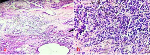
Figure 2. Immunohistochemical staining a: pAkt showed cytoplasmic expression in oral cancer b:pAkt expression was negative in oral cancer c: Inflammatory lesion did not express pAkt.
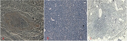
Table 1. pAkt expression pattern and its association with clinicopathological findings.
Figure 3. Immunohistochemical staining a: VEGF showed cytoplasmic expression in oral cancer b: VEGF expression was negative in oral cancer c: Inflammatory lesion did not show any expression.
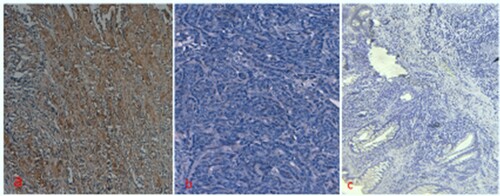
Table 2. VEGF expression pattern and its association with clinical parameters of oral cancer cases.
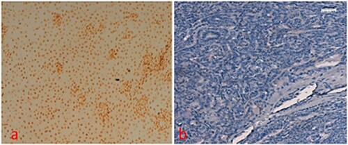
Figure 4. TUNEL staining a: apoptosis seen in oral cancer b: apoptosis expression was negative in oral cancer.