
Figures & data
Table 1. Sequences of primers used for RT-qPCR.
Figure 1. TGF-β1 promoted the proliferation and migration of hASMC. Cells were treated with 10 ng/mL TGF-β1 for 24 h. (A) The cell viability was measured by CCK8 assay. (B and C) The cell migration was assessed by transwell assay. Data are expressed as mean ± SD (n = 3). ***p < 0.001 and ****p < 0.0001 vs vehicle.
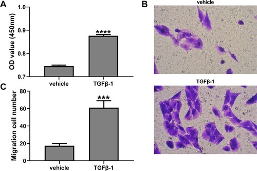
Figure 2. SXD inhibited TGF-β1-induced proliferation and migration of hASMC. Cells were pretreated with different concentrations of SXD (0.1, 0.2 and 0.5 mg/mL) for 6 h, and then treated with or without 10 ng/mL TGF-β1 for 24 h. (A) Cell viability was detected by CCK8 assay. (B and C) Cell migration was assessed by transwell assay. Data are expressed as mean ± SD (n = 3). ****p < 0.0001 vs. control; #p < 0.05, ##p < 0.01, ###p < 0.001 vs. TGF-β1; SXD, Six-ingredient-Xiao-qing-long decoction.
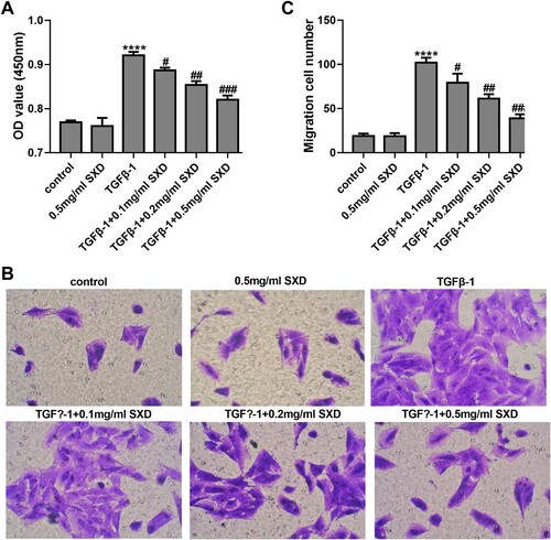
Figure 3. TGF-β1 inhibited FKBP51 expression in hASMC. Cells were treated with 10 ng/mL TGF-β1. (A) The FKBP51 mRNA level was detected by RT-qPCR assay. (B) The FKBP51 protein level was detected by western blotting. Data are expressed as mean ± SD (n = 3). **p < 0.01, ***p < 0.001 vs. 0 h.
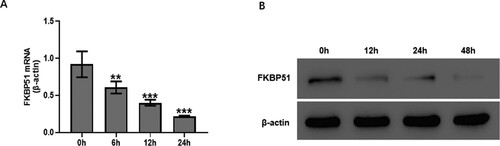
Figure 4. SXD attenuated TGF-β1-mediated FKBP51 expression and AKT phosphorylation. Cells were pretreated with different concentrations of SXD (0.1, 0.2 and 0.5 mg/mL) for 6 h, and then treated with 10 ng/mL TGF-β1 for 24 h. The protein levels of FKBP51, AKT, p-AKT were measured by western blotting. Data are expressed as mean ± SD (n = 3). SXD, Six-ingredient-Xiao-qing-long decoction.
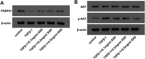
Figure 5. SXD inhibited TGF-β1-induced cell migration by regulating FKBP51/AKT signaling. Cells were transfected with oeFKBP51 or vector. After 24 h of transfection, cells were treated using 10 ng/mL TGF-β1 with or without 0.5 mg/mL SXD for 24 h. (A and B) Cell migration was assessed by transwell assay. (C) The protein levels of FKBP51, AKT and p-AKT were measured by western blotting. Data are expressed as mean ± SD (n = 3). **p < 0.01 vs control; #p < 0.05, ##p < 0.01 vs TGF-β1; SXD, Six-ingredient-Xiao-qing-long decoction.
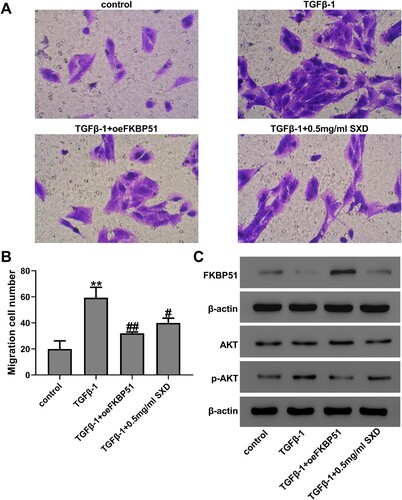
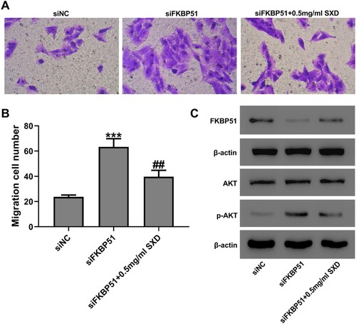
Figure 6. SXD inhibited siFKBP51-induced hASMC migration. Cells were transfected with siFKBP51 or siNC and then cultured with or without 0.5 mg/mL SXD for 24 h. (A and B) Cell migration was assessed by Transwell assay. (C) The protein levels of FKBP51, AKT and p-AKT were measured by western blotting. Data are expressed as mean ± SD (n = 3). ***p < 0.001 vs siNC; ##p < 0.01 vs siFKBP51; SXD, Six-ingredient-Xiao-qing-long decoction.
Supplemental Material
Download Zip (746.6 KB)Data availability statement
Due to the nature of this research, participants of this study did not agree for their data to be shared publicly, so supporting data is not available.