
Figures & data
Figure 1. Dec1 is involved in LPS-induced cell pyroptosis of HPDLFs. A, RT–PCR analysis showing that the expression level of IL-1β was significantly induced by treatment with 10 µg/ml P. gingivalis LPS. B, RT–PCR analysis showing that the expression level of the transcription factor Dec1 was elevated by treatment with LPS. C, Left, Western blot analysis showing that the cleavage of Caspase-1 and GSDMD was activated by LPS, followed by the induction of phosphorylated NF-κB. Right, Quantitation of band density corrected against GAPDH showing means ± SD (n ≥ 3); *p < 0.05, **p < 0.01. All results are representative of at least three independent experiments.
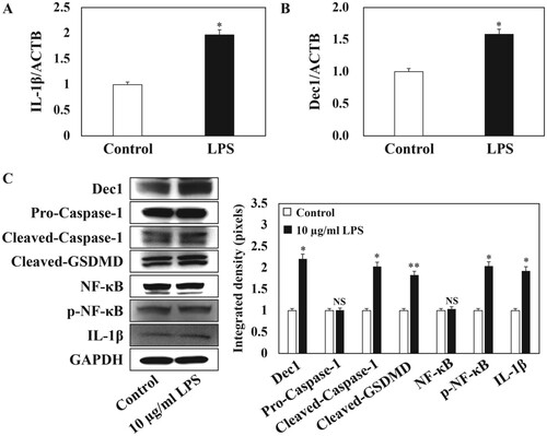
Figure 2. LPS treatment activates Caspase protein expression in HPDLFs. Immunocytofluorescence analysis showing that the expression of Caspase-1 and Caspase-11 was upregulated in the cytoplasm of 10 µg/ml P. gingivalis LPS-induced pyroptosis. Consistently, the downstream protein, p-NF-κB, and Dec1 were also activated and were translocated into the nuclei. The arrows indicate positive cells. Original magnification: 40×, scale bars = 50 µm. All results are representative of at least three independent experiments.
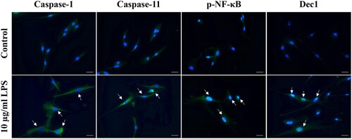
Figure 3. Reduced expression of Dec1 in HPDLFs attenuates LPS-induced pyroptosis. A, RT–PCR analysis showing that the expression of Dec1 was significantly suppressed after Dec1 siRNA transfection with or without LPS treatment. B, RT–PCR analysis showing that the reduced expression of Dec1 attenuated the expression level of IL-1β under LPS induction compared with the control with or without LPS treatment. C, Left, Western blot showing that the inhibition of Dec1 significantly impaired the expression of cleaved-Caspase-1 and cleaved-GSDMD, suppressed the phosphorylation of NF-κB, and decreased IL-1β expression. Right, Quantitation of band density corrected against GAPDH showing means ± SD (n ≥ 3); *p < 0.05, **p < 0.01. All results are representative of at least three independent experiments.
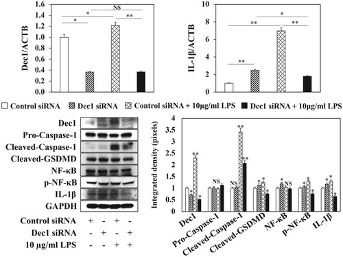
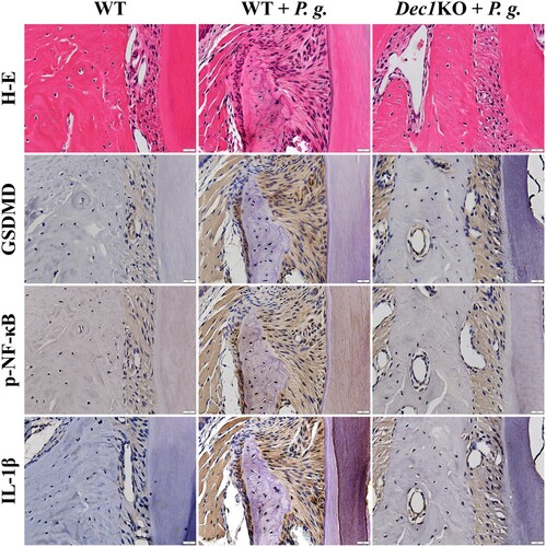
Figure 4. The absence of Dec1 reduces pyroptosis in P. gingivalis-induced periodontitis in mice. Immunohistochemistry showing that the expression of GSDMD, p-NF-κB and IL-1β in the periodontal ligament of WT and Dec1KO mice was suppressed by the absence of Dec1. Original magnification: 60×, scale bars = 20 µm. All results are representative of at least three independent experiments.
Data availability statement
The data that supporting the findings of this study are openly available in figshare at https://doi.org/10.6084/m9.figshare.13102865.v1.