
Figures & data
Table 1. Quantitative phytochemical compositions of ethanol extract of Cassia sieberiana leaves.
Table 2. Effect of EECSL on body weight gain of the rats.
Table 3. Effect of EECSL on the weight of prostate, bladder, testes, and prostate index.
Table 4. Effect of EECSL on serum PSA of testosterone-induced BPH.
Table 5. Effect of EECSL on serum testosterone and DHT levels of testosterone-induced BPH.
Table 6. Effect of EECSL on lipid profile of testosterone-induced BPH.
Table 7. Effect of EECSL on oxidative stress markers in testosterone-induced BPH rats.
Table 8. Effect of EECSL on liver marker enzymes.
Figure 1. Normal control: Photomicrograph of prostate tissue showing normal infoldings (convolutions), corpora amylacea (CA) (red arrow), Luminal secretions (LS) (blue arrow) are adequate and intact seminal vesicles (black arrow). H&E. mag. 400×.
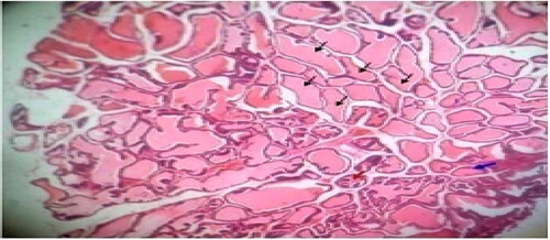
Figure 2. Untreated control: Photomicrograph of prostrate tissue showing slight disruption of architectural appearances (blue arrow) and multiple blood congestions (black star) within the fibromuscular stroma. Enlarged acini (black arrow) also observed. H&E. mag. 400×.
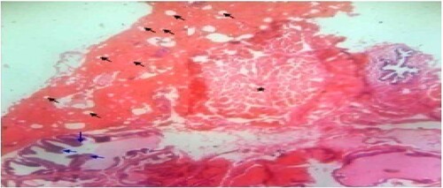
Figure 3. Standard control: Photomicrograph of prostate tissue showed back-to-back micro acini with improved intra acina papillary infoldings/or convolutions (black arrow) of the seminal vesicles duct with fibrous capsules (star). Reduced secretions and focal blood congestions were observed within the fibromuscular stroma of the prostate. H&E. mag. 100×.
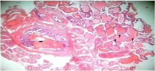
Figure 4. Low dose: Photomicrograph of prostate tissue showed ectasia or dilation of the prostate characterized by acini distended with secretory materials (SM) (star), compression(black arrow) of surrounding unaffected acini with dilation appearing cystic, with fused cuboidal epithelium (blue arrow) lining were present. Infoldings or occasional papillary infoldings was observed. H&E. mag. 400×.
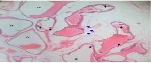
Figure 5. Mid dose: Photomicrograph of dorsal prostate tissue showed extensively enlarged acini with reduced secretory (star) materials in the lumen. The hyperplastic condition (black arrow) was also observed. H&E. mag. 400×.
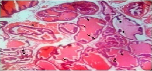
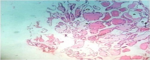
Figure 6. High dose: Photomicrograph of prostate tissue showed glandular hyperplasia predominated and characterized by exaggerated intra-acinar papillary infoldings (convolutions) (black arrow) with fibrovascular cores. Infiltrates mainly lymphocytic cells were observed in the acinar without any admixed tumor cells. Secretions (star) in the acini were observed. H&E. mag. 400×.
Data availability statement
The data that support the findings of this study are available in OSFHOME check finder tool at https://mfr.osf.io/render?url=https%3A%2F%2Fosf.io%2F3grd9%2Fdownload