Figures & data
Figure 1. Aβ caused neurotoxicity and decreased miR-137 level in a dose- and time-dependent manner. (A) MTT assay to assess cell viability following Aβ treatment or control vehicle treatment (Con). (B) GluA1 level in neurons following Aβ treatment or control vehicle treatment (Con). (C) Quantification of (B). (D) Relative Caspase 3 activity in neurons following Aβ treatment or control vehicle treatment (Con). (E) Relative miR-137 level in neurons following Aβ treatment or control vehicle treatment (Con). (n = 4). One-way ANOVA post-hoc Bonferroni. **P < 0.01. (Comparisons were done between groups at same time point but with different concentrations). #P < 0.05; ###P < 0.001 (Comparisons were done between groups at different time points but with same concentrations).
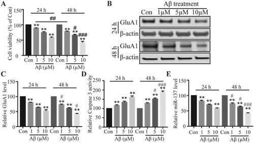
Figure 2. Overexpression of miR-137 reduced the neurotoxicity caused by Aβ. (A) Relative miR-137 level in neurons transfected with mimic-NC (NC) or miR-137 mimics. Student’s t-test. (B) Viability of neurons transfected mimic-NC or miR-137 mimics neurons following Aβ treatment (5 μM for 48 h) compared with control vehicle treated neurons (Con). (C, D) GluA1 protein level in transfected neurons following Aβ treatment (5 μM for 48 h) compared with control vehicle treated neurons (Con). One-way ANOVA post-hoc Bonferroni. (E) Relative Caspase 3 activity in transfected neurons following Aβ treatment (5 μM for 48 h) compared with control vehicle treated neurons (Con). One-way ANOVA post-hoc Bonferroni. (n = 4) ** P < 0.01.
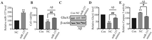
Figure 3. miR-137 directly targeted ERK1/2. (A) Complementary binding sites between miR-137 and ERK1/2 mRNA. (B) Relative luciferase activity of WT-ERK1/2 and MUT-ERK1/2 in transfected neurons. Student’s t-test (n = 4). (C, D) ERK1/2 protein level in neurons transfected with mimic-NC or miR-137 mimics. Student’s t-test. (n = 4) n.s., not significant; ** P < 0.01.
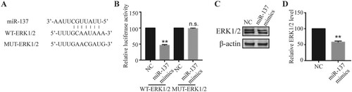
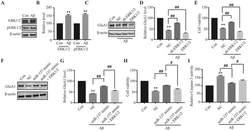
Figure 4. miR-137 protected neurons from Aβ-induced neurotoxicity via targeting ERK1/2. (A, B) ERK1/2 and pERK1/2 protein levels in neurons with Aβ treatment (5 μM for 48 h) compared with control vehicle treated neurons (Con). Student’s t-test. (C, D) GluA1 level in neurons transfected with NC, sh-ERK1/2, or ERK1/2 following Aβ treatment (5 μM for 48 h) compared with control vehicle treated neurons (Con). One-way ANOVA post-hoc Bonferroni. (E) Relative viabilities of neurons transfected with NC, sh-ERK1/2, or ERK1/2 following Aβ treatment (5 μM for 48 h) compared with control vehicle treated neurons (Con). One-way ANOVA post-hoc Bonferroni. (F, G) GluA1 level in neurons transfected with NC, sh-ERK1/2, or ERK1/2 following Aβ treatment (5 μM for 48 h) compared with control vehicle treated neurons (Con). One-way ANOVA post-hoc Bonferroni. (H) Relative viabilities of neurons transfected with NC, sh-ERK1/2, or ERK1/2 following Aβ treatment (5 μM for 48 h) compared with control vehicle treated neurons (Con). One-way ANOVA post-hoc Bonferroni. (I) Relative Caspase 3 activity in neurons transfected with NC, sh-ERK1/2, or ERK1/2 following Aβ treatment (5 μM for 48 h) compared with control vehicle treated neurons (Con). One-way ANOVA post-hoc Bonferroni. (n = 4). **P < 0.01; #P < 0.05; ##P < 0.01.