
Figures & data
Figure 1. Chemical structure of hirsutenone molecule.
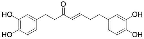
Figure 2. (A) Results of MTT assay. Thyroid MDA-T32 cancer cells were treated with hirsutenone at varying indicated doses for 24, 48 and 72 h. It was observed that the viability of target cells reduced in a dose as well as time-dependent manner. (B) MTT assay results of the effect of hirsutenone on the viability of normal thyroid cells namely follicular epithelial cell line-Nthy-ori 3-1. Results indicated that normal cells showed lesser cytotoxicity as compared to the cancer cells. Data is shown as means ± standard deviation with repetitions n = 3. *P < 0.05.
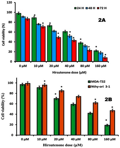
Figure 3. (A) Pictures representing thyroid MDA-T32 cancer cell colonies after 48 h of hirsutenone treatment and 10 days of incubation. Experiments were performed three times. (B) Graphical representation of number of cell colonies after hirsutenone exposure. It was observed that number of cell colonies appeared to be decreasing with increasing drug doses. Data is revealed as means ± standard deviation with replications n = 3. *P < 0.05.
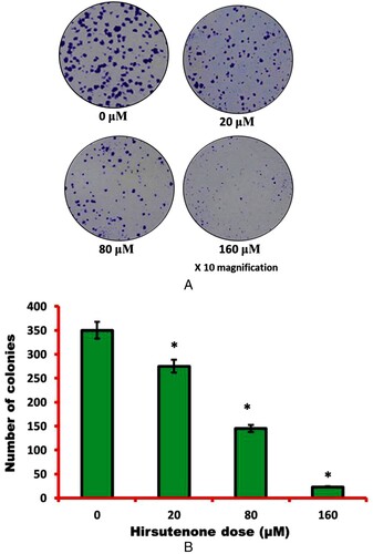
Figure 4. To examine cell morphology of hirsutenone treated thyroid MDA-T32 cancer cells, AO/EB staining assay was performed. Results revealed apoptotic cell morphology as represented by arrows like nuclear condensation, DNA fragmentation and membrane blebbing.
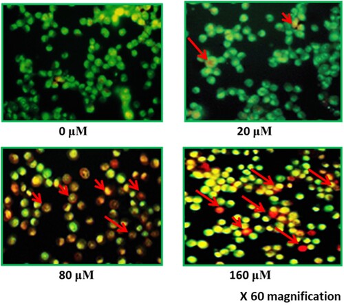
Figure 5. (A) Flow cytometric analysis of hirsutenone treated MDA-T32 cells after performing annexin V/PI staining assay. Results representing increasing number of apoptotic cell percentage with increasing drug doses. Data is shown as means ± standard deviation with replications n = 3. P < 0.05. (B)0 Graphical representation of increased number of apoptotic cells after execution of annexin V/PI staining assay at indicated doses. Data is shown as means ± standard deviation with replications n = 3. *P < 0.05.
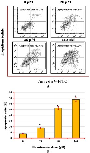
Figure 6. Results representing the activity of pro and anti-apoptotic proteins including caspases (−3, −8 and −9). Experiments were repeated three times.
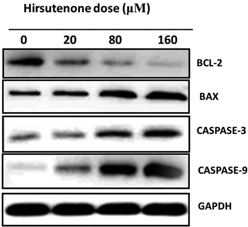
Figure 7. Wound healing assay results presenting wound closure width at controls and hirsutenone treated MDA-T32 cells at indicated doses. Experiments were repeated three times.
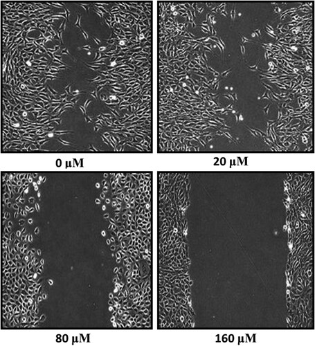
Figure 8. Transwell chambers assay featuring reduced rate of invasion after hirsutenone treatment at indicated doses. Experiments were repeated three times.
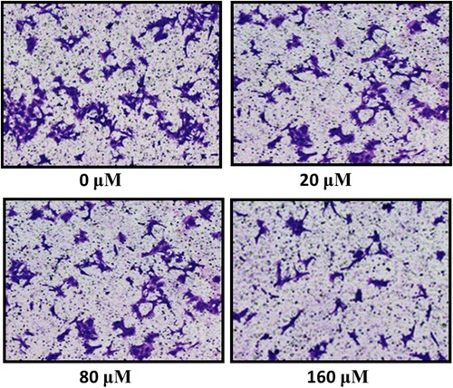
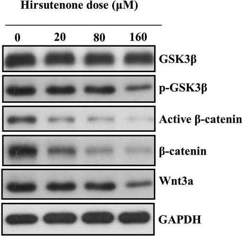
Figure 9. Protein expression of Wnt/beta-catenin signalling pathway associated proteins after MDA-T32 cells were exposed to indicated concentrations of hirsutenone drug. Individual experiments were repeated three times.
Data availability statement
The data which was generated and analysed during current study are always available from the corresponding author and on request will be made available. The data is freely available from the figshare repository (https://figshare.com/) and can be easily accessed at: https://figshare.com/articles/dataset/Antiproliferative_effects_of_Hirsutenone_in_thyroid_cancer_cells/12780818.