
Figures & data
Figure 1. Levels of BNP and MMP10 in the three groups of rats The normal control group reported lower levels of BNP (A) and MMP10 (B) compared with the other groups (all P < 0.05). Both BNP and MMP10 were lower in the DCM + treatment group than the DCM + control group (& P < 0.05).
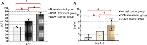
Table 1. Levels of BNP and MMP10 in the three groups of rats (±s).
Table 2. Qualitative indexes of ventricular remodeling in the three groups of rats (±s).
Figure 2. Comparison of heart weight index (HWI) and left ventricular weight index (LVWI) in the three experimental groups Both HWI and LVWI were lower in the normal control group than in the DCM + treatment and DCM + control groups (*P < 0.05 for all).
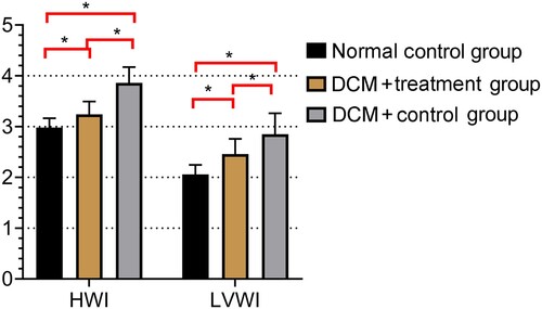
Table 3. BW, HWI, and LVWI of the three groups of rats (±s).
Table 4. Effects of astragalus heart-protecting decoction on the levels of oxidative stress markers in the three groups of rats (±s).
Figure 3. Changes in myocardial histology in the three groups of rats HE staining (×200) showing the pathomorphology of the myocardial tissues in the three groups of rats. (A) Normal histology in the myocardial tissues of the rats in the normal control group. (B) Cardiomyocyte necrosis was observed under the intima of the left ventricle and throughout the myocardium in the DCM + control group. (C) Tissues of rats in the DCM + treatment group exhibited less severe necrotic fibrosis, a small amount of fibrosis adjacent to some cardiomyocytes, and better cardiomyocyte arrangement.
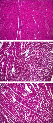
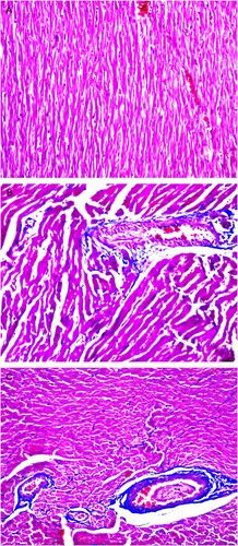
Figure 4. Degree of myocardial fibrosis in the three groups of rats Masson staining (×200) showing the degree of myocardial fibrosis in the three groups of rats, Erythrocytes, cardiac muscle fibers, and cytoplasm appear red, whereas myocardial interstitial collagen appears blue. (A) Masson staining mainly showing normal myocardial tissues in the normal control group. (B) Myocardial interstitial collagen fiber accumulation and obvious hyperplasia were observed in the myocardium of the DCM + control group. (C) Blue-stained collagen fibers were observed in the myocardium of the DCM + treatment group.
Data availability statement
The data that support the findings of this study are available in figshare at http://doi.org/10.6084/m9.figshare.15112242