1,739
Views
2
CrossRef citations to date
0
Altmetric
Cancer Biology

Inhibition of autolysosomes by repurposing drugs as a promising therapeutic strategy for the treatment of cancers

Pages 568-601 | Received 29 Sep 2021, Accepted 02 Mar 2022, Published online: 24 May 2022

Figures & data

Table 1. Dysfunctional autophagy is associated with different human diseases.

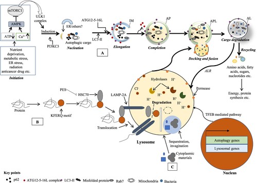
Figure 1. Different classes of autophagy process. The autophagy is classified as (A) macroautophagy where cytosolic constituents are submerged and delivered to the lysosome; (B) microautophagy where cytosolic wastage products are straightly submerged and co-opted by lysosome via redisposition of its membrane and (C) chaperone-mediated autophagy (CMA) where KFERQ motif holding cellular proteins documented by the cytosolic HSP70 chaperone and translocated to the lysosomal membrane via interacting with LAMP2 receptor leading to their degradation. Macroautophagy (simply autophagy) proceeds the isolation membrane (IM) formation. Cellular stresses culminate in AMPK stimulation and mTORC1 suppression that in turn makes the ULK1 complex dissociated with mTORC1 and initiates autophagy. Through a series of interchain responses, this IM confiscates its cargos into an autophagosome (AP, a double-membrane vesicle) that in turn kisses and fuses with the lysosome to make autolysosome (AL). The AL cargos are degraded by an acidic niche containing hydrolytic enzymes in lysosomal lumen. The degrading products are used for energy production or biosynthesis of macromolecules. TFEB coordinates the stress responses like starvation via lysosome and controls countless cellular processes including autophagy and lysosomal biogenesis (Al-Bari and Xu Citation2020).

Figure 1. Different classes of autophagy process. The autophagy is classified as (A) macroautophagy where cytosolic constituents are submerged and delivered to the lysosome; (B) microautophagy where cytosolic wastage products are straightly submerged and co-opted by lysosome via redisposition of its membrane and (C) chaperone-mediated autophagy (CMA) where KFERQ motif holding cellular proteins documented by the cytosolic HSP70 chaperone and translocated to the lysosomal membrane via interacting with LAMP2 receptor leading to their degradation. Macroautophagy (simply autophagy) proceeds the isolation membrane (IM) formation. Cellular stresses culminate in AMPK stimulation and mTORC1 suppression that in turn makes the ULK1 complex dissociated with mTORC1 and initiates autophagy. Through a series of interchain responses, this IM confiscates its cargos into an autophagosome (AP, a double-membrane vesicle) that in turn kisses and fuses with the lysosome to make autolysosome (AL). The AL cargos are degraded by an acidic niche containing hydrolytic enzymes in lysosomal lumen. The degrading products are used for energy production or biosynthesis of macromolecules. TFEB coordinates the stress responses like starvation via lysosome and controls countless cellular processes including autophagy and lysosomal biogenesis (Al-Bari and Xu Citation2020).

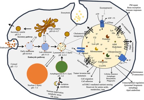
Figure 2. The central position of lysosomes at the ALP. Activated lysosomes and autolysosomes are associated with terminal degradation stage through endocytic and autophagic pathways. Exogenous products are endocytosed by endosomes and autolysosomes act as an integral part of the autophagy for degradation. The membrane of lysosome plays as a podium for the gathering of mTORC1 complex, which plays a main role for nutrient sensing signals of lysosomes to control energy levels through downstream mediators including TFEB. Interior pHs of intracellular vesicles steadily decrease laterally from early endosomes to late endosomes to lysosomes, the mostly acidic organelles. Lysosomes create and preserve their pH homeostasis by applying v-ATPase carrier. The positive charged ions such as K+ can be dispatched by K+ ion channel or by exchanged by Cl influx carrier via ClC-7, a Cl/H+ antiporter.

Figure 2. The central position of lysosomes at the ALP. Activated lysosomes and autolysosomes are associated with terminal degradation stage through endocytic and autophagic pathways. Exogenous products are endocytosed by endosomes and autolysosomes act as an integral part of the autophagy for degradation. The membrane of lysosome plays as a podium for the gathering of mTORC1 complex, which plays a main role for nutrient sensing signals of lysosomes to control energy levels through downstream mediators including TFEB. Interior pHs of intracellular vesicles steadily decrease laterally from early endosomes to late endosomes to lysosomes, the mostly acidic organelles. Lysosomes create and preserve their pH homeostasis by applying v-ATPase carrier. The positive charged ions such as K+ can be dispatched by K+ ion channel or by exchanged by Cl− influx carrier via ClC-7, a Cl−/H+ antiporter.

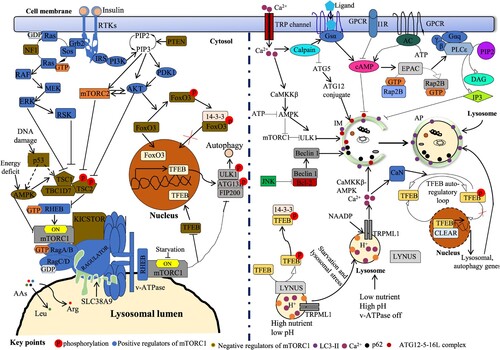
Figure 3. Control of ALP by mTORC1-dependent and independent signal transductions. Amino acid (AA) signal, growth factors like insulin and cellular ATP regulate autophagy via mTORC1 mTORC1-dependent and independent pathways. Insulin stimulates both mTORC1 and mTORC2 via the traditional PI3K-AKT-TSC1/2 (tuberous sclerosis complex 1/2)-RHEB pathway. In Ras-MAPK (ERK) signal, ERK and RSK suppress TSC2 to induce mTORC1. Low nutrition status induces AMPK and TSC complex, suppressing RHEB and mTORC1. Carriage of AAs controls mTORC1 action via AA transporters like SLC3A2 in cell membrane as well as SLC38A9 in lysosomal membrane. The RAGULATOR complex ties up the RAGs to the surface of lysosome. V-ATPase is essential for the induction of mTORC1 on lysosomal surface. RAG A/B is controlled by the GATOR1 complex and GATOR2. KICSTOR complex acts as an essential action in tethering GATOR1 to the lysosomal surface, thus suppressing the activity of RAG A/B and mTORC1. Lysosomal membrane-oriented arginine (Arg) transporter, SLC38A9 binds with the RAGs and RAGULATOR to stimulate mTORC1. DNA damage inhibits mTORC1 activity by activating p53 in an AMPK- and TSC1/2-conditional way. Activated mTORC1 controls TFEB intracellular position via its phosphorylation (at Ser211) and subsequent confiscation with 14-3-3 protein. mTORC2 regulates autophagy via FOXO3 pathway. Phosphorylated FOXO3 interacts with 14-3-3 protein in cytoplasm and prevents induction of gene transcription. mTOR-independent cell signaling controlling autophagy comprises cAMP-Epac-PLCϵ-IP3, Ca2+-calpains–Gsα and inositol (Ins) signaling pathways. Intracytosolic Ca2+ status can be amplified by multiple Ca2+ channels including the TRP channel. An upsurge in cytosolic Ca2+ levels stimulates calpains and then Gsα/AC/cAMP/Epac/Rap2B/PLCϵ signal which regulates autophagy by endorsing IP3 via its receptor, IP3R activation. IP3 binds to IP3 receptor (IP3R) on ER to release stored Ca2+ that impairs AP maturation by blocking AP-lysosome fusion. Ca2+ efflux through lysosomal NAADP triggered TRP channel can be extra augmented by Ca2+ secretion from IP3R causing activation of the CaMKKβ–AMPK signal and AP formation. However, Ca2+ secretion from lysosomes can influence lysosomal pH and repeal fusion of lysosomes with APs. On the other hand, an upsurge Ca2+ concentration in cytosol next stimulates CAMKK-β, tracked by AMPK-mediated autophagy induction. In normal cases, phosphorylated TFEB by mTORC1 is sequestered with 14-3-3 protein in cytoplasm. Under fasting, lysosomal mTORC1 is inactive form and upsurge Ca2+ is secreted from lysosome via TRPML1 channel. Then Ca2+ stimulates CaN (calcineurin) that interacts and dephosphorylates TFEB. This form of TFEB is incapable to interact with 14-3-3 protein and easily moves to the nucleus (Al-Bari and Xu Citation2020).

Figure 3. Control of ALP by mTORC1-dependent and independent signal transductions. Amino acid (AA) signal, growth factors like insulin and cellular ATP regulate autophagy via mTORC1 mTORC1-dependent and independent pathways. Insulin stimulates both mTORC1 and mTORC2 via the traditional PI3K-AKT-TSC1/2 (tuberous sclerosis complex 1/2)-RHEB pathway. In Ras-MAPK (ERK) signal, ERK and RSK suppress TSC2 to induce mTORC1. Low nutrition status induces AMPK and TSC complex, suppressing RHEB and mTORC1. Carriage of AAs controls mTORC1 action via AA transporters like SLC3A2 in cell membrane as well as SLC38A9 in lysosomal membrane. The RAGULATOR complex ties up the RAGs to the surface of lysosome. V-ATPase is essential for the induction of mTORC1 on lysosomal surface. RAG A/B is controlled by the GATOR1 complex and GATOR2. KICSTOR complex acts as an essential action in tethering GATOR1 to the lysosomal surface, thus suppressing the activity of RAG A/B and mTORC1. Lysosomal membrane-oriented arginine (Arg) transporter, SLC38A9 binds with the RAGs and RAGULATOR to stimulate mTORC1. DNA damage inhibits mTORC1 activity by activating p53 in an AMPK- and TSC1/2-conditional way. Activated mTORC1 controls TFEB intracellular position via its phosphorylation (at Ser211) and subsequent confiscation with 14-3-3 protein. mTORC2 regulates autophagy via FOXO3 pathway. Phosphorylated FOXO3 interacts with 14-3-3 protein in cytoplasm and prevents induction of gene transcription. mTOR-independent cell signaling controlling autophagy comprises cAMP-Epac-PLCϵ-IP3, Ca2+-calpains–Gsα and inositol (Ins) signaling pathways. Intracytosolic Ca2+ status can be amplified by multiple Ca2+ channels including the TRP channel. An upsurge in cytosolic Ca2+ levels stimulates calpains and then Gsα/AC/cAMP/Epac/Rap2B/PLCϵ signal which regulates autophagy by endorsing IP3 via its receptor, IP3R activation. IP3 binds to IP3 receptor (IP3R) on ER to release stored Ca2+ that impairs AP maturation by blocking AP-lysosome fusion. Ca2+ efflux through lysosomal NAADP triggered TRP channel can be extra augmented by Ca2+ secretion from IP3R causing activation of the CaMKKβ–AMPK signal and AP formation. However, Ca2+ secretion from lysosomes can influence lysosomal pH and repeal fusion of lysosomes with APs. On the other hand, an upsurge Ca2+ concentration in cytosol next stimulates CAMKK-β, tracked by AMPK-mediated autophagy induction. In normal cases, phosphorylated TFEB by mTORC1 is sequestered with 14-3-3 protein in cytoplasm. Under fasting, lysosomal mTORC1 is inactive form and upsurge Ca2+ is secreted from lysosome via TRPML1 channel. Then Ca2+ stimulates CaN (calcineurin) that interacts and dephosphorylates TFEB. This form of TFEB is incapable to interact with 14-3-3 protein and easily moves to the nucleus (Al-Bari and Xu Citation2020).

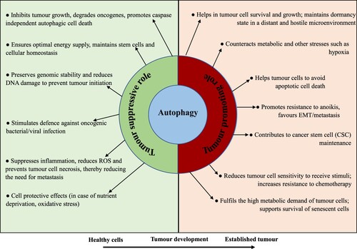
Figure 4. The Janus-faced contradictory actions of autophagy in cancer cells. Autophagy acts as a Janus-faced play or the Yin-Yang faced function on cancer development. In healthy cells, it functions as a tumor inhibitory mechanism by ensuring optimal energy supply, preserving genomic integrity, promoting the degradation of damaged organelle and cellular oncogenes, and endorsing first-line immunity against microbial infection. In advanced tumors, however, autophagy actions can be reinstated. Autophagy functions as an oncogenic phenomenon by endorsing cancer development via declining stresses, the preservation of cancer stemness potential and metastasis; executing cells resistant to anoikis; helping the persistence of cancer cells in a dormant state and supporting the senescent cell state.

Figure 4. The Janus-faced contradictory actions of autophagy in cancer cells. Autophagy acts as a Janus-faced play or the Yin-Yang faced function on cancer development. In healthy cells, it functions as a tumor inhibitory mechanism by ensuring optimal energy supply, preserving genomic integrity, promoting the degradation of damaged organelle and cellular oncogenes, and endorsing first-line immunity against microbial infection. In advanced tumors, however, autophagy actions can be reinstated. Autophagy functions as an oncogenic phenomenon by endorsing cancer development via declining stresses, the preservation of cancer stemness potential and metastasis; executing cells resistant to anoikis; helping the persistence of cancer cells in a dormant state and supporting the senescent cell state.

Table 2. Selected examples of ALP inhibitors with therapeutic benefits.

Table 3. Selected examples of therapeutic benefits from ALP inhibition in animal models.

Table 4. Ongoing and completed clinical trials with ALP inhibitors.
Data availability statement

Data sharing does not apply to this article as no new data were created or analyzed in this study.