
Figures & data
Figure 1. Structure of Skin membrane barrier showing different layers of skin tissue like stratum corneum followed by epidermis and lastly dermis along with different routes of permeation through skin.
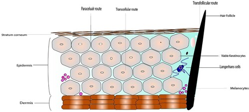
Figure 2. Classification of Skin cancer.
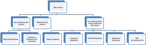
Figure 3. Different types of Nanocarriers used in topical/dermal therapy.

Table 1. Different types of research studies showing performance of different nanoparticle system in skin cancer treatment with major emphasis given to 5-fluorouracil with some studies with different API.
Figure 4. Various nanocarriers of different classes – 2a- Lipid based nanocarriers (Liposome), 2b- Polymer based nanocarriers (Polymeric micelle) 2c- Inorganic nanocarriers (Gold nanoparticle)-1479 with permission from Springer Nature - License no: 5010250744068).
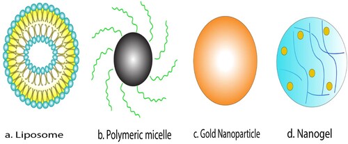
Figure 5. TEM images showing the uniform size of 20 nm of Silica NPs (Adapted from Lee et al. Citation2016. A quantitative study of nanoparticle skin penetration with interactive segmentation. Medical & biological engineering & computing, 54(10), pp.1469-1479 with permission from Springer Nature – License no: 5010250744068).
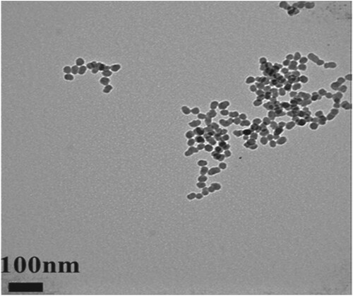
Figure 6. Negative charge image (Adapted from Lee et al. Citation2016. A quantitative study of nanoparticle skin penetration with interactive segmentation. Medical & biological engineering & computing, 54(10), pp.1469-1479 with permission from Springer Nature - License no: 5010250744068)
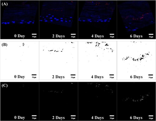
Figure 7. Positive charge image (Adapted from Lee et al. Citation2016. A quantitative study of nanoparticle skin penetration with interactive segmentation. Medical & biological engineering & computing, 54(10), pp.1469-1479 with permission from Springer Nature - License no: 5010250744068)
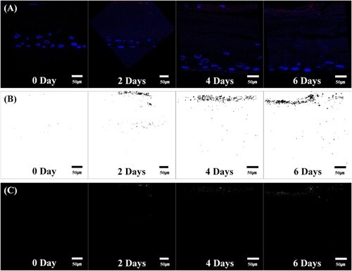
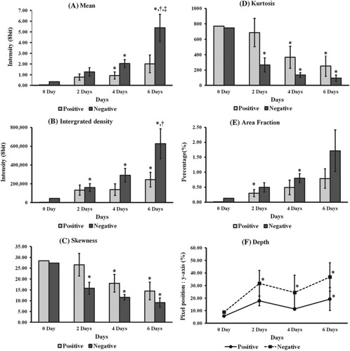
Figure 8. 7 Quantification results for each group. (A) Mean, (B) integrated density, (C) skewness, (D) kurtosis, (E) area fraction, (F) penetration depth. *vs. Day 0 Group, †vs. Day 2 Group, ‡vs. Day 4 Group (Student's t test, P < 0.05).
Data availability statement
Data sharing is not applicable to this article as no new data were created or analysed in this study.