
Figures & data
Table 1. Details of the participants.
Figure 1. (A) occlusal view of the sensor sheet attached to the palate, (B) positioning of surface electrodes over the orofacial muscles.
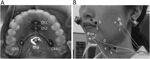
Figure 2. A typical example of tongue pressure waveforms, raw and rectified waveforms of orofacial muscle activities during swallowing.
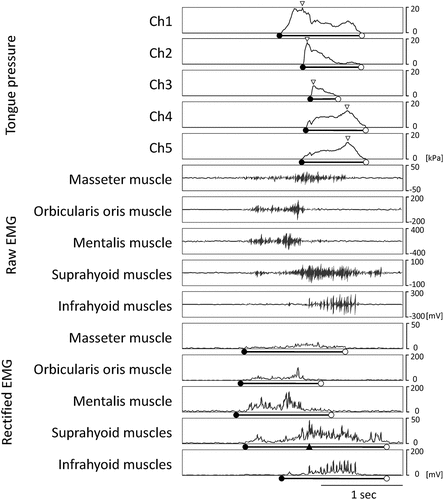
Figure 3. Assessment of palatal morphology on the plaster model of the maxilla.
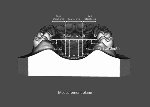
Figure 4. (A) time sequences for tongue pressure and orofacial muscle activities, (B) duration of tongue pressure and orofacial muscle activities, (C) maximum magnitude of tongue pressure.
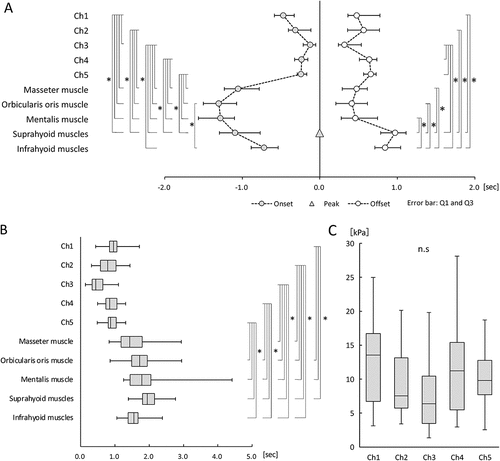
Figure 5. Palatal depth.
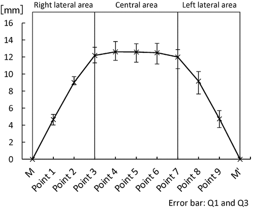
Table 2. Analysis of palatal morphology.
Table 3. Relationships between the maximum magnitude of tongue pressure and palatal morphology.
Table 4. Relationships of the onset of tongue pressure production and orofacial muscle activities with palatal morphology.
Table 5. Relationships of the offset of tongue pressure production and orofacial muscle activities with palatal morphology.
Table 6. Relationships of the duration of tongue pressure production and orofacial muscle activities with palatal morphology.