Figures & data
Figure 1. Preparation of polyacrylic acid (PAA) nanoparticles through reverse micellar polymerization.
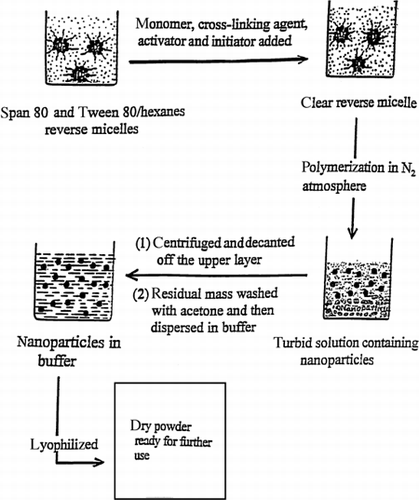
Figure 2. Quasielastic laser light scattering by PAA nanoparticles in water.
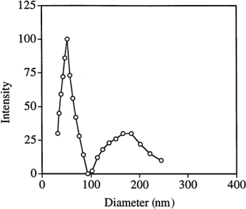
Figure 3. TEM micrographs of PAA nanoparticles.
Table 1. Polyacrylic Acid Particle Sizes in Water as Measured by Quasielastic Laser Light Scattering
Figure 4. Quasielastic laser light scattering by PAA nanoparticles in phosphate buffer (50 mM, pH 7.0). Particle sizes are shown before and after lyophilization.
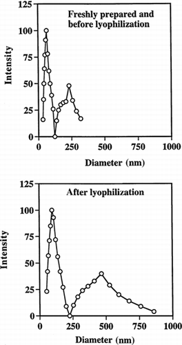
Figure 5. Quasielastic laser light scattering by PAA nanoparticles in phosphate buffer (50 mM, pH 7.0). Particles sizes are shown before and after storage.
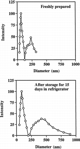
Figure 6. Loading timolol maleate (TM) into PAA nanoparticles as a function of TM concentration in the loading solution. (MBA is the crosslinker.)
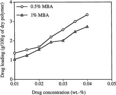
Figure 7. Release of TM into deionized water through dialysis tubing. (np signifies nanoparticles.)
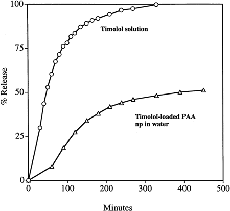
Figure 8. Release of timolol maleate (TM) by dialysis into deionized water or phosphate buffer (50 mM phosphate, pH 7.4). Data are shown for a TM-loaded PAA nanoparticulate (np) suspension, or a TM solution, each as separate dialysis experiments.
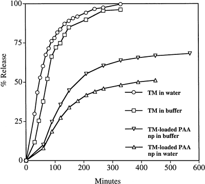
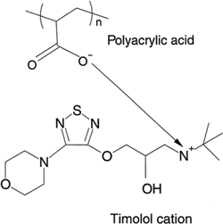
Figure 9. Structural formula of the timolol cation and the polyacrylic acid anion, demonstrating possible ionic bonding between the two.