Figures & data
Figure 1. Effect of PEG-HbV on agonist-induced platelet aggregation. Platelets were treated without (open circle) or with (closed circle) 5.8 mg/dl (Hb) of PEG-HbV at 37°C for 3 min on an aggregometer prior to addition of various concentrations of collagen, thrombin or ristocetin. Values are means ± SE of three donors. Error bars are not shown when smaller than the symbols.
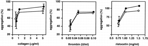
Figure 2. Effect of PEG-HbV on collagen-induced RANTES release from washed platelets. Washed platelets were incubated with various concentrations of PEG-HbV at 37°C for 10 min and then stimulated without (open column) or with (hatched column) collagen at 37°C for 5 min. The levels of RANTES in the supernatant of the mixture were measured by ELISA. Values are means ± SE of five donors. *p < 0.05, in comparison with control (no PEG-HbV)
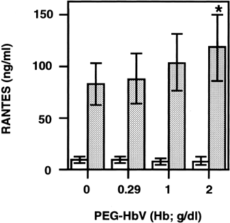
Figure 3. Effect of PEG-HbV on collagen-induced RANTES release from PRP. Platelets were incubated with various concentrations of PEG-HbV at 37°C for 10 min and then stimulated without (open column) or with (hatched column) collagen for 5 min at 37°C. The concentration of collagen used was 1 μg/ml in (A) and 2 μg/ml in (B). Values are means ± SE of five donors (A) or three donors (B).
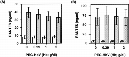
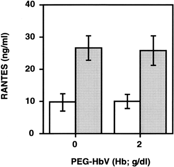
Figure 4. Effect of longer incubation of PRP with PEG-HbV on collagen-induced RANTES release. Platelets were incubated with or without 2 g/dl (Hb) of PEG-HbV at 37°C for 60 min, and then stimulated without (open column) or with 1 μg/ml of collagen (hatched column). Values are means ± SE of five donors.