334
Views
25
CrossRef citations to date
0
Altmetric
Original

LOW-POWER DIODE LASER STIMULATION OF SURGICAL OSTEOCHONDRAL DEFECTS: RESULTS AFTER 24 WEEKS

, , , , , & show all
Pages 235-244 | Published online: 11 Jul 2009

Figures & data

Table I. Scoring System for Histologic and Morphometric Results (Maximum = 12 Points). Samples (n.6 per Group) Were Prepared 24 Weeks After Surgery

Figure 1. Osteochondral defect at 24 weeks after laser treatment (toluidine blue). Magnification: ×10. Complete healing of the osteochondral defect surgically created; the arrow shows the border between repaired tissue (right) and adjacent tissue (left). The repaired tissue has an overall good structural integrity with a smooth and regular articular surface.
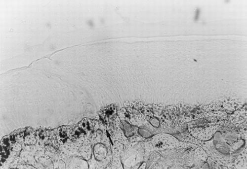
Figure 1. Osteochondral defect at 24 weeks after laser treatment (toluidine blue). Magnification: ×10. Complete healing of the osteochondral defect surgically created; the arrow shows the border between repaired tissue (right) and adjacent tissue (left). The repaired tissue has an overall good structural integrity with a smooth and regular articular surface.

Table II. Mean Cell Density ± SD of the Lased Specimens. Each Sample Is the Result of the Comparison Between the Cartilage Repaired Using the Laser and the Cartilage Adjacent to the Lesion

Figure 2. Osteochondral defect at 24 weeks after laser treatment (safranin-0 and picrosirius red). Magnification: ×20. Cartilaginous cells appear arranged in a palisade structure; matrix staining of the reconstituted cartilage is more intense for both proteoglycans and collagen in the deepest layers.
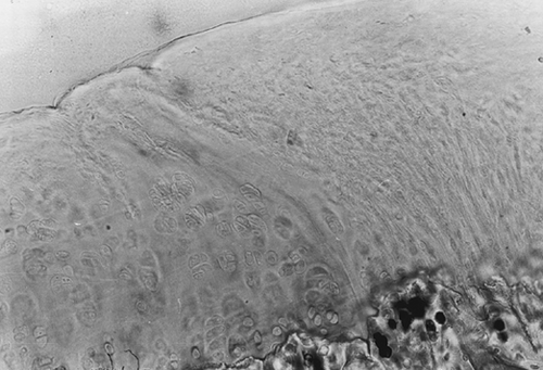
Figure 2. Osteochondral defect at 24 weeks after laser treatment (safranin-0 and picrosirius red). Magnification: ×20. Cartilaginous cells appear arranged in a palisade structure; matrix staining of the reconstituted cartilage is more intense for both proteoglycans and collagen in the deepest layers.

Figure 3. Osteochondral defect at 24 weeks without laser treatment (toluidine blue). Magnification: ×2.5. The repaired tissue appears disorganized and reveals an irregular growth of the reconstituted bony tissue, a thin layer of fibrous tissue on the articular surface and the absence of chondrocytes.
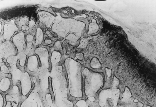
Figure 3. Osteochondral defect at 24 weeks without laser treatment (toluidine blue). Magnification: ×2.5. The repaired tissue appears disorganized and reveals an irregular growth of the reconstituted bony tissue, a thin layer of fibrous tissue on the articular surface and the absence of chondrocytes.

Reprints and Corporate Permissions

Please note: Selecting permissions does not provide access to the full text of the article, please see our help page How do I view content?

To request a reprint or corporate permissions for this article, please click on the relevant link below:

Academic Permissions

Please note: Selecting permissions does not provide access to the full text of the article, please see our help page How do I view content?

Obtain permissions instantly via Rightslink by clicking on the button below:

If you are unable to obtain permissions via Rightslink, please complete and submit this Permissions form. For more information, please visit our Permissions help page.