Figures & data
Figure 1. Interaction of rGPIa/IIa-Ibα-liposomes with platelets on the collagen surface depends on the shear rate. Images were obtained after a 3-min perfusion of a mixture of liposomes (4.0 × 10⁵/μl) and platelets (1.0 × 10⁵/μl) on the surface at a hematocrit of 37.5%, soluble vWf concentration of 10 μg/ml, 2 mM Mg2+, 37°C, and various shear rates as indicated. Exofacial concentration of rGPIa/IIa and rGPIbα was 1.0 and 0.70 μg/ml, respectively. Values are the mean ± standard deviation, n = 6.
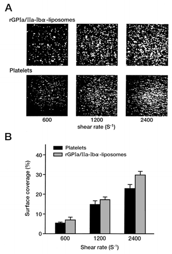
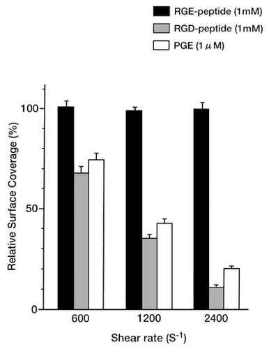
Figure 2. Inhibition of the interaction of rGPIa/IIa-Ibα-liposomes with platelets on the collagen surface. Experimental conditions were the same as described for . Inhibition studies were performed in the presence of the RGD-peptide (1 mM), RGE-peptide (1 mM), or PGE (1 μM).
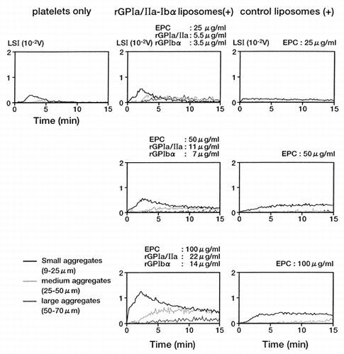
Figure 3. Enhancement of platelet aggregation by rGPIa/IIa-Ibα-liposomes assessed by changes in light scattering intensity. PRP at 1 × 10⁴/μl platelets was mixed with various concentrations of rGPIa/IIa-Ibα-liposomes, and 10 μg/ml of collagen was then added to the mixture.
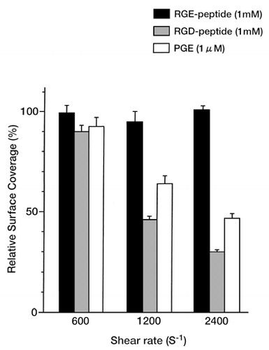
Figure 4. Interaction of Fbg-liposomes with platelets on the collagen surface depends on the shear rate. Experimental conditions were the same as described for , except that the exofacial concentration of Fbg was 0.74 μg/ml.
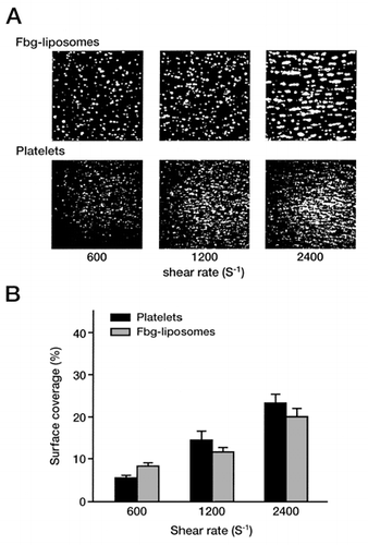
Figure 5. Inhibition of the interaction of Fbg-liposomes with platelets on the collagen surface. Experimental conditions were the same as described for .
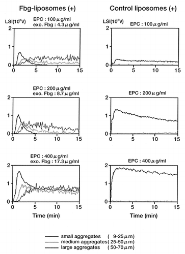
Figure 6. Enhancement of platelet aggregation by Fbg-liposomes assessed by changes in light scattering intensity. PRP at 5 × 10⁴/μl platelets was mixed with various concentrations of Fbg-liposomes, and 2 μM ADP was then added to the mixture.