Figures & data
Figure 1. Molecular distribution of cross-linked and non–cross-linked ultrapure bovine SF-Hb. Elution profiles of 5-, 10-, 24-hour cross-linked PolyHb, PolyHb-SOD-CAT and non–cross-linked bovine SF-Hb were obtained by running 1 ml of the 10× diluted samples on Sephadex G-200 1.6 cm×70 cm column (VT=105.5 ml, Vo=38 ml, equilibrated with 0.1 M Tris-HCl, pH 7.5, and eluted at 11.5 ml/hr).
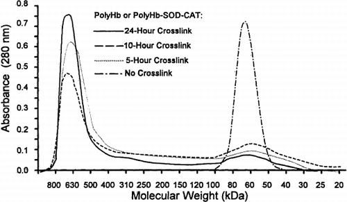
Table 1. Molecular Components of PolyHb and PolyHb-SOD-CAT
Table 2. Molecular Distribution and SOD and CAT Activity
Figure 2. Oxygen-hemoglobin dissociation. The 24 hours of cross-linking with glutaraldehyde did not significantly affect the P50 value of SF-Hb. PolyHb and PolyHb-SOD-CAT P50 values were 26.1±0.2 mmHg and 24.7±0.2 mmHg, respectively. The SF-Hb P50 value was 27.8±0.0 mmHg. All oxygen-hemoglobin dissociation curves are presented as averages of 6 trials (n=6).
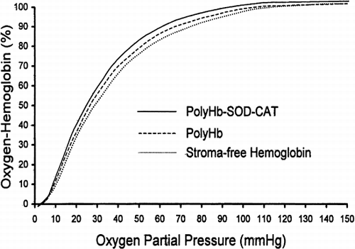
Figure 3. Determining the required ischemic duration. Animals were subjected to either 0, 20, 30, 40, or 60 minutes of ischemia before reperfusion. With 60 minutes of ischemia, the level of Evans blue in the sham control was significantly lower than that of oxygenated saline, stroma-free Hb (SF-Hb), and PolyHb. Statistical significance is P<0.01.
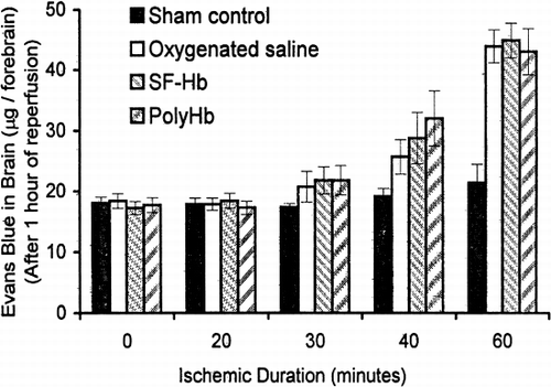
Figure 4. Evans blue extravasation. PolyHb-SOD-CAT significantly attenuated the severity of BBB disruption as compared to saline, SF-Hb, SF-Hb+SOD+CAT, and PolyHb. Statistical significance is P<0.01.
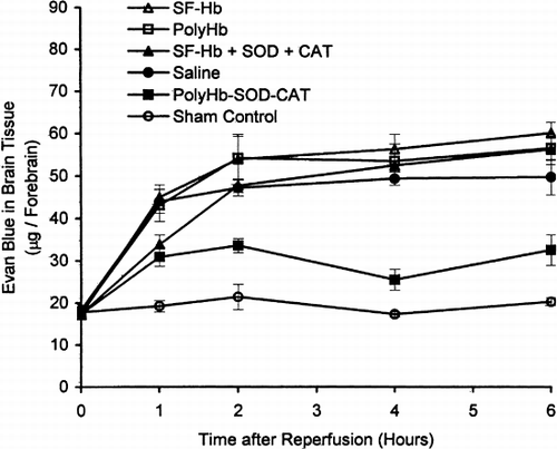
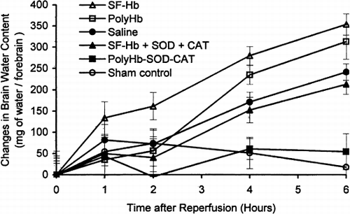
Figure 5. Brain edema: changes in brain water content. The changes in brain water content of PolyHb-SOD-CAT–treated animals were not significantly different from that of the sham control. The increase in water contents of saline, SF-Hb, SF-Hb+SOD+CAT, and PolyHb were significantly different from that of the sham control and PolyHb-SOD-CAT group by the 4th hour and increased with time. Statistical significance is P<0.01.