Figures & data
Figure 1. Hepatocyte microcapsules made by single-step encapsulation method; some cells could be seen entrapped in the capsular membrane (arrows).
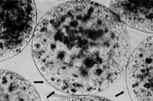
Figure 2. (a) Gel beads formed in the 100 mM CaCl2 in the first step of the two-step encapsulation method. Each large bead usually contains 3 small beads. (b) Microcapsules made by two-step encapsulation, after the inner small beads solubilize, the capsular membrane is uniform, no cells are seen entrapped in the membrane.
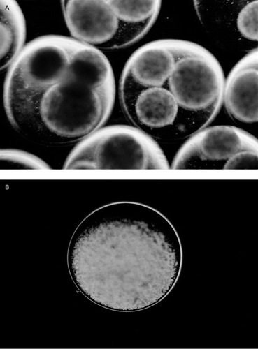
Figure 3. (a) Recovered microcapsules at week 4 in Group I, these hepatocytes microcapsules were made by single-step encapsulation method. The microcapsules were attached to the greater omentum and connective tissue. (b) The recovered microcapsules in a were mechanically destroyed and stained by trypan blue. The capsular membrane was infiltrated with lymphocytes and macrophages.
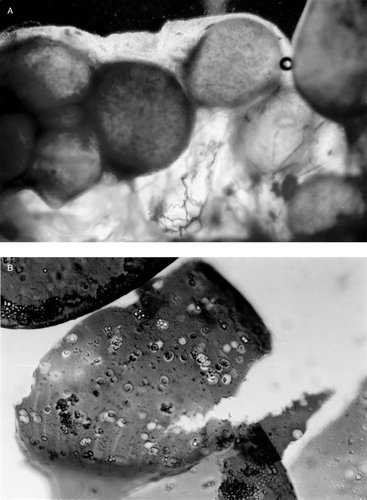
Figure 4. Recovered microcapsules made by two-step encapsulation method, at week 6 in Group IV (hepatocytes and bone marrow cells co-encapsulation). These capsular membranes were smooth; no inflammatory cells or macrophage infiltrated the membrane.
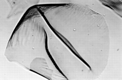
Figure 5. (a) Group IV (hepatocytes and bone marrow cells co-encapsulation by two-step method). At week 6, the recovered microcapsules were disrupted mechanically to release the content. The hepatocytes released were in an aggregated form. (b) After using Trypsin–EDTA, the aggregated cells in recovered microcapsules were dispersed and the hepatocyte viability could be determined.
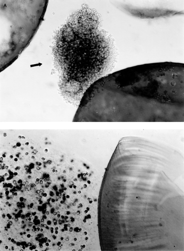
Figure 6. Hepatocytes viability in recovered microcapsules in Group I and Group II. Group I: Single-step encapsulation hepatocytes microcapsules. Group II: Single-step encapsulation hepatocytes and bone marrow cells co-microcapsules. There was no significant difference of recovered hepatocytes viability up to week 2 post-implantation. *From week 3, microcapsules were infiltrated with fibrous tissue and the viability of hepatocytes could not be determined. The values are mean±SD in three rats.
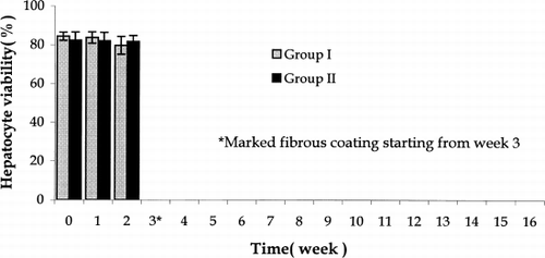
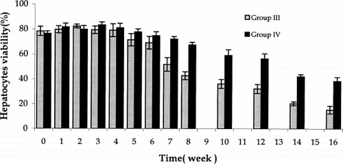
Figure 7. Hepatocytes viability in recovered microcapsules in Group III and Group IV. Group III: Two-step encapsulation hepatocytes microcapsules. Group IV: two-step encapsulation hepatocytes and bone marrow cells co-microcapsules. In Groups III and IV, from week 7 to week 16, the hepatocytes viability in Group IV was significantly higher than in Group III. The values are means±SD in three rats.