Figures & data
Figure 1. Schematic of computational domain with subregions RBC, plasma, vascular wall, interstitium and tissue.
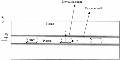
Table 1. List of Model Parameters
Figure 2. Comparison of ko, for different initial PO2. P50,cHb=P50,sHb=29.3 Torr, nc=ns=2.2, [Hb]s=7g/dl, Hc=0.2.
![Figure 2. Comparison of ko, for different initial PO2. P50,cHb=P50,sHb=29.3 Torr, nc=ns=2.2, [Hb]s=7g/dl, Hc=0.2.](/cms/asset/6b8827f4-80cd-4666-94b0-eed44020bb02/ianb19_a_11116809_uf0002_b.gif)
Figure 3. (A) Comparison of k o for different values of HBOC oxygen affinities. P 50,cHb=29.3 Torr, nc=ns=2.2. Hc=0.2, [Hb] s=7 g/dl. (B) Comparison of kcell for different values of HBOC oxygen affinities. P50,cHb=29.3 Torr, nc=ns=2.2, Hc=0.2, [Hb] s=7 g/dl.
![Figure 3. (A) Comparison of k o for different values of HBOC oxygen affinities. P 50,cHb=29.3 Torr, nc=ns=2.2. Hc=0.2, [Hb] s=7 g/dl. (B) Comparison of kcell for different values of HBOC oxygen affinities. P50,cHb=29.3 Torr, nc=ns=2.2, Hc=0.2, [Hb] s=7 g/dl.](/cms/asset/715303b8-5e9c-4344-814e-eca5a460aa1c/ianb19_a_11116809_uf0003_b.gif)
Figure 4. (A) Comparison of k o for different values of HBOC oxygen cooperatives. P 50,c Hb=P 50,s Hb=29.3 Torr, n c=2.2, H c=0.2, [Hb] s=7 g/dl. (B) Comparison of k cell for different values of HBOC oxygen cooperatives. P 50,c Hb=P 50,s Hb=29.3 Torr, n c=2.2, H c=0.2, [Hb] s=7 g/dl.
![Figure 4. (A) Comparison of k o for different values of HBOC oxygen cooperatives. P 50,c Hb=P 50,s Hb=29.3 Torr, n c=2.2, H c=0.2, [Hb] s=7 g/dl. (B) Comparison of k cell for different values of HBOC oxygen cooperatives. P 50,c Hb=P 50,s Hb=29.3 Torr, n c=2.2, H c=0.2, [Hb] s=7 g/dl.](/cms/asset/2a3deeb3-ab94-45b3-bf1f-a613040c0e4b/ianb19_a_11116809_uf0004_b.gif)
Figure 5. (A) Comparison of k o for different values of HBOC concentrations. P 50,c Hb=P 50,s Hb=29.3 Torr, n c=n s=2.2, H c=0.2. (B) Comparison of k cell for different values of HBOC concentrations. P 50,c Hb=P 50,s Hb=29.3 Torr, n c=n s=2.2, H c=0.2.
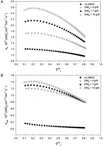
Figure 6. (A) Tissue PO 2 distribution along capillary length for different HBOC oxygen affinities. P 50,c Hb=29.3 Torr, n c=n s=2.2, H c=0.2, [Hb] s=7 g/dl. (B) Radial tissue PO 2 distribution profiles for different HBOC oxygen affinities. P 50,c Hb=29.3 Torr, n c=n s=2.2, H c=0.2, [Hb] s=7 g/dl. The vertical line shows the plasma-wall interface. A-arteriolar end, M-midcap, V-venular end. –·–·–·–· P 50,s Hb=10 Torr, ------- P 50,s Hb=29.3 Torr, ——— P 50,s Hb=50 Torr.
![Figure 6. (A) Tissue PO 2 distribution along capillary length for different HBOC oxygen affinities. P 50,c Hb=29.3 Torr, n c=n s=2.2, H c=0.2, [Hb] s=7 g/dl. (B) Radial tissue PO 2 distribution profiles for different HBOC oxygen affinities. P 50,c Hb=29.3 Torr, n c=n s=2.2, H c=0.2, [Hb] s=7 g/dl. The vertical line shows the plasma-wall interface. A-arteriolar end, M-midcap, V-venular end. –·–·–·–· P 50,s Hb=10 Torr, ------- P 50,s Hb=29.3 Torr, ——— P 50,s Hb=50 Torr.](/cms/asset/4c781fd9-4fcc-47e5-9ee5-a27ccfd73fe3/ianb19_a_11116809_uf0006_b.gif)
Figure 7. Tissue PO 2 distribution along capillary length for different HBOC oxygen cooperatives. P 50,c Hb=P 50,s Hb=29.3 Torr, n c=2.2, H c=0.2, [Hb] s=7 g/dl.
![Figure 7. Tissue PO 2 distribution along capillary length for different HBOC oxygen cooperatives. P 50,c Hb=P 50,s Hb=29.3 Torr, n c=2.2, H c=0.2, [Hb] s=7 g/dl.](/cms/asset/66f39bb3-7f86-4247-bd8f-0c9378b55e35/ianb19_a_11116809_uf0007_b.gif)
Figure 8. Tissue PO 2 distribution along capillary length for different HBOC concentrations. P 50,c Hb=P 50,s Hb=29.3 Torr, n c=n s=2.2, H c=0.2.
Figure 9. (A) Comparison of k o for different values of hematocrits. P 50,c Hb=P 50,s Hb=29.3 Torr, n c=n s=2.2, [Hb] s=7 g/dl. (B) Comparison of k cell for different values of hematocrits. P 50,c Hb=P 50,s Hb=29.3 Torr, n c=n s=2.2, [Hb] s=7 g/dl.
![Figure 9. (A) Comparison of k o for different values of hematocrits. P 50,c Hb=P 50,s Hb=29.3 Torr, n c=n s=2.2, [Hb] s=7 g/dl. (B) Comparison of k cell for different values of hematocrits. P 50,c Hb=P 50,s Hb=29.3 Torr, n c=n s=2.2, [Hb] s=7 g/dl.](/cms/asset/d3fa156a-9946-45c1-a0e2-17a3ffeacda8/ianb19_a_11116809_uf0009_b.gif)
Figure 10. Comparison of kinetic parameter values with and without HBOC. P 50,c Hb=P 50,s Hb=29.3 Torr, n c=n s=2.2, [Hb] s=7 g/dl.
![Figure 10. Comparison of kinetic parameter values with and without HBOC. P 50,c Hb=P 50,s Hb=29.3 Torr, n c=n s=2.2, [Hb] s=7 g/dl.](/cms/asset/198ea137-9f16-486f-8e96-f8c0362a08f4/ianb19_a_11116809_uf0010_b.gif)
Figure 11. Comparison of overall mass transfer coefficient for different HBOC diffusion coefficients. P 50,c Hb=P 50,s Hb=29.3 Torr, n c=n s=2.2, [Hb] s=7 g/dl.
![Figure 11. Comparison of overall mass transfer coefficient for different HBOC diffusion coefficients. P 50,c Hb=P 50,s Hb=29.3 Torr, n c=n s=2.2, [Hb] s=7 g/dl.](/cms/asset/56c7b240-bd72-4e47-b205-2deb97d55a6e/ianb19_a_11116809_uf0011_b.gif)